Self-healable PEDOT-based film, hydrogel, and OECT devices for bioelectronics
Introduction
In recent years, bioelectronics has made remarkable progress in developing wearable and implantable devices that can monitor and interact with biological systems due to the development of conducting polymer-based self-healing materials.1 The human body is composed of living organisms characterized by the presence of bioelectricity, which is produced by the flow of ions across biological membranes. Measuring these electrical signals allows for the evaluation of different physiological parameters of the body, such as the electroencephalogram (EEG) for the brain, the electrocardiogram (ECG) for the heart, and the electromyography (EMG) for muscle responses.2 With its customized and non-invasive healthcare options, bioelectronics promises to transform medical practices, providing new ways to diagnose and treat various illnesses.3
Materials used in bioelectronics are required to mimic the properties of biological tissues. In this context, the current devices made of rigid inorganic materials may experience damage or degradation from physical stress and chemical interactions. On the other hand, soft organic polymers combine mechanical flexibility and high electrical conductivity, allowing for the development of more advanced bioelectronics.4 Another challenge in bioelectronics is the requirement of conductive materials to maintain their performance for extended periods. Self-healing conductors have gained attention due to their ability to repair damages in their conducting structure, thus enhancing the durability and longevity of electronic devices by restoring electrical conductivity. This makes them a potential solution to address the issue at hand. Self-healing can occur through various mechanisms, including chemical reactions, physical interactions (mechanical or thermal), or shape-memory effects.5 In bioelectronics, self-healing materials are beneficial as they can repair themselves in dynamic biological environments.6
The organic conducting polymer, poly(3,4-ethylenedioxythiophene) (PEDOT) doped with polystyrene sulfonate (PSS), has been widely used for developing self-healable conductors due to its ease of processing in aqueous media, high electrical conductivity, and self- healing properties.1 Herein, we discuss the recent advances in self- healable conductors based on PEDOT:PSS (Cat. Nos. 483095, 900208, 900181) and organic electrochemical transistor (OECT) devices based on these materials.
Self-healable PEDOT:PSS-based Films
The conjugated structure of organic conducting polymers, composed of carbon atoms with alternating single and double bonds along the polymer backbone, facilitates electron delocalization along the entire chain and leads to electrical conductivity. The conductivity of the conjugated system can be enhanced by doping, such as PSS- doped PEDOT.7 In the PEDOT:PSS system, the conductivity can be further improved by incorporating conductivity enhancers, such as glycerol (Cat. No. G7893), sorbitol (Cat. No. 56755-M), ethylene glycol (EG, Cat. No. 102466), and dimethyl sulfoxide (DMSO, Cat. No. 276855).8 Pure conducting polymers generally exhibit poor mechanical properties, characterized by brittleness and fragility, making them susceptible to mechanical damage such as cracking or breaking. Therefore, they must be combined with other materials to improve their mechanical properties. Self-healing conductive materials have been obtained by blending conducting polymers with other polymers, such as polyvinyl alcohol (PVA, Cat. No. 341584), polyethylene glycol (PEG, Cat. No. 202398), Triton™ X-100 (Cat. No. X100), agarose, and others. In the next section, we will introduce the self-healing performance and mechanical properties of PEDOT:PS-based materials.
Our group has reported that PEDOT:PSS film can undergo water- induced healing.9 In this work, the PEDOT:PSS films were drop-cast onto a glass slide, and the current flow was monitored throughout the process. The current flow was interrupted when a cut was made intentionally with a razor blade. The current was recovered after a water droplet was placed on the damaged area (Figure 1A). The cutting and healing process was repeated in various regions of the film, and excellent healing efficiency of 100% and rapid response time of 150 ms were observed. The scanning electron microscopy (SEM) image of the damaged PEDOT:PSS film reveals a noticeable cut gap before water contact and mechanical gap recovery after healing (Figure 1B). Adding water to the separated PEDOT:PSS films may cause them to swell and increase in volume, enabling the filling of wounded areas and the restoration of conductivity. Based on the observation of the water-enabled rapid healing property of PEDOT:PSS thin films, wireless water sensors using water-healable PEDOT:PSS films were demonstrated.
In a successive work, we investigated the water-induced healing property of PEDOT:PSS-based films by studying the impact of 3-(glycidyloxypropyl)trimethoxysilane (GOPS, Cat. No. 440167) incorporation and the effect of sulfuric acid post-treatment.10 The healing performance can be tailored by adjusting the concentration of GOPS and the duration of sulfuric acid treatment. Increasing the volume ratio of GOPS to PEDOT:PSS from 0.5 to 1 resulted in a conductivity decrease from 350 S cm-1 to 300 S cm-1 and a healing efficiency reduction from 66% to 15%. While sulfuric acid post- treatment significantly improved the conductivity of PEDOT:PSS films, the healing efficiency decreased to 93%, 75%, and 70%, depending on the soaking time in strong acid (Figure 1E). It is reported that increasing GOPS content and sulfuric acid treatment duration influences the swelling of PSS, which was observed as a decrease in weight gain of the PEDOT:PSS films, resulting in deterioration of self-healing ability. In addition, we also explored PEDOT films with different dopants. We observed that inserting organic dopants, tosylate (Tos) and triflate (OTf), through chemical polymerization can lead to water-enabled healing properties. In contrast, the inorganic perchlorate ion (ClO4-) doped film obtained from electro-polymerization showed no healing ability.
When blended with a small amount (<10%) of certain additives, the PEDOT:PSS-based films exhibit autonomous healing without needing assistance from water. This exceptional capability is achieved by adjusting the film’s mechanical properties with these additives.11 This proposed strategy enhances the autonomous self- healing property typically observed only in wet PEDOT:PSS film. PEDOT:PSS mixed with low molecular weight PEG (200 or 400) have achieved autonomous healing and showed 100% healing efficiency with a 1 second response time (Figure 1C). With an increase in PEG molecular weight to 1500, the healing efficiency decreased to 80% (Figure 1C). We reported that the high molecular weight of PEG caused lower chain mobility, resulting in decreased viscoelastic behavior in the film. Adding PEG 400 to PEDOT:PSS decreased storage and loss modulus while increasing the elongation at break from 1% to 9%. Therefore, the composite exhibited viscoelastic behavior, and autonomous healing was achieved at a PEG concentration of 4 vol%.
Adding Triton X-100 to PEDOT:PSS can improve its viscoelastic properties, allowing for repetitive electrical current healing without external stimulation (Figure 1D). This film has been reported to have a stable electrical response during repetitive folding and stretching.12 The polymer blend of PEDOT:PSS and a tandem repeat protein derived from squid ring teeth (SRT) has exhibited electrical and mechanical healing abilities at 70 °C in wet conditions. The recovery was observed when 40 wt% SRT was added to PEDOT:PSS due to the interaction between PSS and SRT proteins at 70°C in the presence of water.13
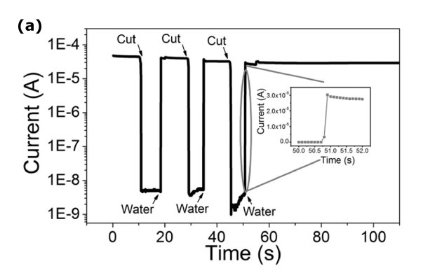


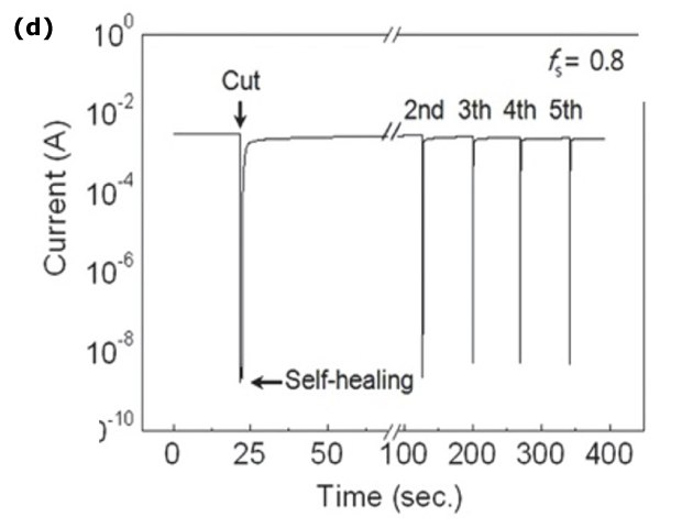
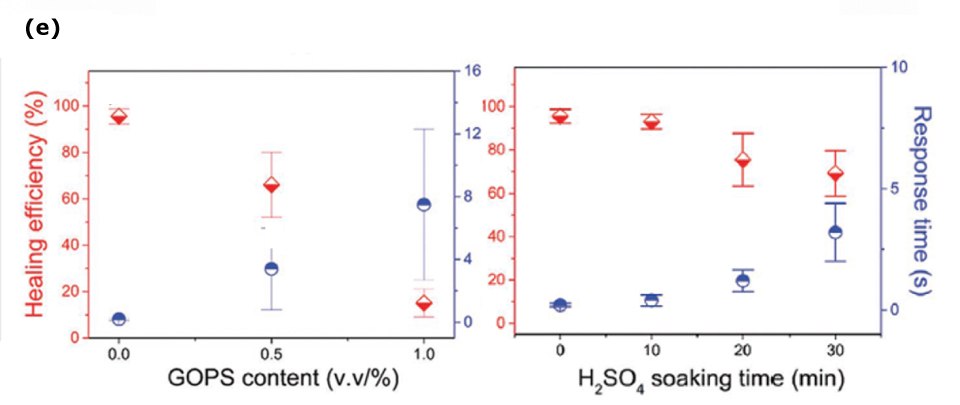
Figure 1. A) Current vs. time measurement demonstrating water-induced electrical healing of PEDOT:PSS film. B) SEM images of the damaged and healed areas of PEDOT:PSS film. Reprinted with permission from reference 9, copyright 2017 Wiley Online Library. C) Current vs. time measurement with several cuts showing the different electrical healing capabilities of PEDOT:PSS films with 4% PEG-400, PEG-200, and PEG-1500. Reprinted with permission from reference 11, copyright 2020 Wiley Online Library. D) Current vs. time test on PEDOT:PSS/Triton X-100 system revealing the healing property. Reprinted with permission from reference 12, copyright 2016 Wiley Online Library. E) Healing efficiency and response time versus different amounts of GOPS and sulfuric acid immersion time of PEDOT:PSS films. Reprinted with permission from reference 10, copyright 2020 Wiley Online Library.
Self-healable Hydrogels
Conductive and self-healable hydrogels show great potential in bioelectronics and wearable electronics owing to their tunable mechanical and electrical properties, high water content, biocompatibility, and self-healing ability. Generally, self-healable hydrogels can be achieved either by utilizing pure conductive polymers (e.g., PEDOT:PSS) or, in most cases, by incorporating conductive materials (e.g., PEDOT:PSS, silver nanowires) into a matrix of self-healable polymers (e.g., PVA, poly(acrylamide) (PAAM), and gelatin).1
Our group reported self-healable PEDOT: PSS-based hydrogels obtained from mixing PVA, borax, and PEDOT:PSS screen printing ink (CleviosTM SV3, containing PEDOT:PSS, propylene glycol (PG, Cat. No. P4347), and diethylene glycol (DEG, Cat. No. H26456) (Figure 2A).14In this work, the as-prepared SV3/PVA hydrogels exhibited high adhesion (~2 N/cm2 on porcine skin), high plastic stretchability (>10000% strain), and a low compressive Young’s modulus (~4 kPa). In addition, the hydrogels present repeated self-healing abilities with a healing efficiency of ~100% due to the introduction of the ink into the PVA-borax system (Figure 2B). Epidermal electrodes fabricated using the PEDOT: PSS-based self- healing hydrogel exhibited excellent signal quality for ECG and EMG recording, which is comparable to that of commercial Ag/AgCl gel electrodes (Cat. Nos. BASMF2056, BASMF2052).14
The conductive hydrogels have been reported to be prone to environmental factors such as high or low temperatures, which impedes their practical application for long-term use. This is primarily due to water loss, which can adversely affect their mechanical and electrical properties. Water-retaining and anti- freezing organohydrogels have been developed to address this issue by incorporating anti-freezing agents, such as EG and glycerol, into the formulations.15–17 Self-healable organohydrogels obtained from PEDOT:PSS, PVA, and EG showed anti-freezing ability and high stretchability (200–900% strain). In addition, the gels exhibited self-healing behavior with a healing efficiency of ~85% (Figure 2C), which was attributed to the dynamic dissociation and re-association of crystalline domains and hydrogen bonds.18


Figure 2.A) Illustration of SV3/PVA hydrogel preparation. B) Current vs. time plots upon several cutting-healing cycles for the SV3/PVA hydrogel showing repeated electrical self-healing. Reprinted with permission from reference 14, copyright 2022 Elsevier. C) Tensile stress vs. strain plots of the original and healed organohydrogels. Reprinted with permission from reference 16, copyright 2017 Wiley Online Library.
Self-healable OECTs
OECTs, or three-terminal devices, have attracted increasing interest in bioelectronics. An OECT is comprised of source and drain electrodes connected by an ion-permeable conducting polymer channel (e.g., PEDOT:PSS) and a liquid or gel electrolyte in direct contact with the channel and a gate electrode. Although the electrodes are typically prepared by microfabrication, etching, and metal deposition,19 printing is a valuable alternative, especially for flexible and stretchable substrates.20,21 The conducting polymer channel can be deposited via various techniques, including spin coating, electrospinning, vapor phase polymerization, electropolymerization, and printing.22 Concerning the gating media, although aqueous electrolytes are commonly used, gel electrolytes, such as iongels and hydrogels prepared by spin coating or printing, have been explored owing to their promising potential in flexible and stretchable electronics.19,20 The operation of OECT utilizes the interaction between ions from the electrolyte and the channel material. This is achieved by controlling the gate voltage, which drives ions in/out of the channel and alters the doping state of the conducting polymer, leading to a change in the channel conductivity. Gating through an electrolyte allows the OECT to operate under 1 V with large amplification of signals owing to their high volumetric capacitance. The combination of these merits makes OECTs a suitable candidate for biological applications.22
OECTs have been explored to record electrophysiological signals in vivo for healthcare monitoring. For example, ECG signals can be easily acquired by putting an OECT on the skin. Furthermore, the amplified signals of electrooculography (EOG), EEG, and EMG can be recorded to monitor human eye movement, brain activity, and muscle movement.23 OECTs can also be directly interfaced invasively with organs to access their local signals. For example, PEDOT:PSS OECTs have been implanted in rat brains to record epileptic seizures, demonstrating the capability of detecting the signals from a single neuron and inducing localized stimulation by current injection in vivo. Further mapping of the brain was realized by employing soft OECT arrays.24 Another vital application, OECT transducers, performing as ultra-sensitive biosensors, have been broadly investigated to detect ions and metabolites, such as Na+, K+, glucose, and lactate. High sensitivity has been demonstrated in detecting dopamine, adrenaline, DNA (deoxyribonucleic acid), and bacteria.25
Self-healing materials have been integrated into various functional devices, including physical or chemical sensors, organic field- effect transistors, and energy storage devices. However, research on self-healing OECTs and their applications is still limited due to the lack of self-healing channel materials. Developing self-healable materials that are capable of maintaining their charge transport properties simultaneously remains a challenge.26
We reported a ground-breaking study on the first self-healing PEDOT:PSS OECTs. The devices exhibited remarkable healing properties, as evidenced by the preservation of transfer characteristics upon in-situ razor blade damage. This significant finding further highlights the potential of OECTs for long-term bioelectronic applications.9 Subsequent studies explored all- solid-state self-healing OECTs, incorporating surfactant-modified PEDOT:PSS channels and PVA hydrogel electrolytes. The device showed excellent performance with high transconductance, fast response, long-term stability, and ion-sensing behaviors (Figure 3A), making it an interesting candidate for developing practical bioelectronic applications.27 Additionally, PEDOT:PSS hydrogel fibers through syringe injection were investigated as a potential channel material for OECTs and demonstrated self-healing properties, suggesting promising potential for healable bioelectronics.28 Recent work has successfully combined the outstanding stretchability and self-healing properties of PEDOT:PSS films by adding a soft polymer, leading to the successful realization of an OECT array (Figure 3B). This development marks a significant advancement in the field of bioelectronic devices.29 Alternatively, incorporating hydrogel electrolytes has also been shown to endow OECTs with self-healing properties and has been demonstrated to mimic various synaptic functions.30
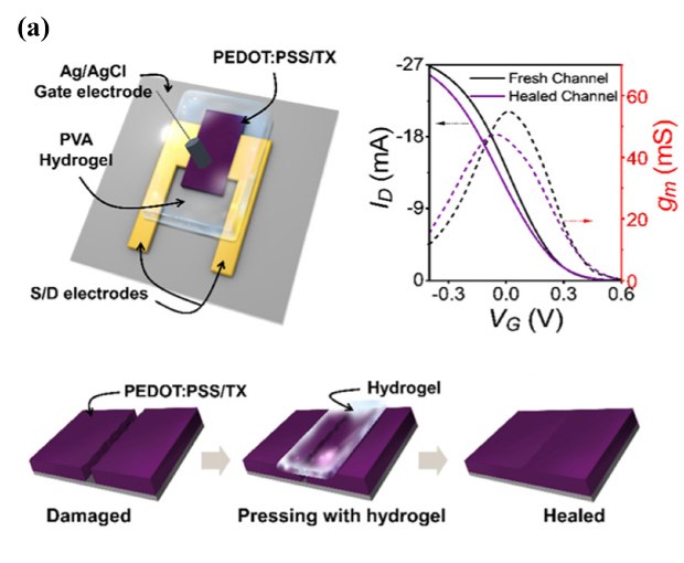
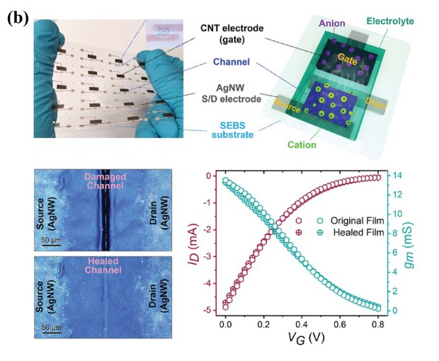
Figure 3. A) Schematic diagram of PEDOT:PSS-based OECT with self-healable performance shown in transfer curve and self-healing process of channel film with the hydrogel on top. Reprinted with permission from reference 22, copyright 2020 American Chemical Society. B) Stretchable and self-healable PEDOT:PSS-based OECTs arrays showing gap recovery and transistor performance restoration. Reprinted with permission from reference 24, copyright 2022 Wiley Online Library.
Conclusion and Outlook
PEDOT:PSS has been demonstrated to be a self-healable conducting polymer. Self-healing PEDOT composite-based films, hydrogels, and devices, mainly OECTs, have been widely explored for various bioelectronic applications. Although significant progress has been achieved in materials development and application discovery, there are still challenges and new chances for self-healable bioelectronics. From the materials perspective, the available conducting polymers with self-healing properties remain limited. Response time and efficiency are two principal metrics when considering the healing properties. Improved performance with rapid recovery and high efficiency are always needed. Furthermore, long-term stability for healing function does face challenges, especially in the case of hydrogels with a high water content that is easily evaporated.
Additionally, due to a lack of electronic materials, research on self-healing electronic devices, or OECTs, is still restricted. The mismatching between layers makes it challenging to create fully healable OECTs with all self-healing layers. In spite of these challenges, a self-healing integrated electronic platform with multicomponents is expected.
References
如要继续阅读,请登录或创建帐户。
暂无帐户?