Utilization of TrueGel3D™ Hydrogels to Study Fibroblast Spreading
Introduction
Fibroblasts that are present in the stroma secrete glycoproteins and polysaccharides to form extracellular matrix. They maintain the structural integrity within the connective tissue and play a key role in cancer, chronic obstructive pulmonary diseases, idiopathic pulmonary fibrosis, scleroderma and several other diseases.
Fibroblasts grown in 3D environments express natural cell physiology closer to in-vivo conditions than their counterpart cultured in 2D environments. TrueGel3D™ hydrogel is a robust, chemically defined hydrogel made up of biologically inert, synthetic biopolymers that can be customized to match the native cell environment. The objective of this study is to investigate the fibroblast spreading within TrueGel3D™ hydrogel when crosslinked with CD cell-degradable and PEG non cell-degradable crosslinkers.
Methods
TrueGel3D™ hydrogel preparation:
All steps were performed in a sterile hood and the volume ratio of each component was added as indicated below.
- 3T3 fibroblasts cell suspension was prepared in culture medium, D-PBS sterile without Ca++/Mg++) or any other physiological solution.
- Water, 10X buffer (pH 5.5) and FAST-PVA were mixed in a reaction tube.
- TrueGel3D™ RGD integrin adhesion peptide was added to the reaction tube containing FAST-PVA and mixed immediately to ensure homogenous distribution followed by incubation for 5 min to allow attachment of the peptide to the maleimide groups of FAST-PVA polymer.
- 3.0 µL of PEG non cell-degradable crosslinker OR CD cell-degradable crosslinker was pipetted and spotted on the surface of a sterile culture plate (8-chambers slide) compatible with inverse microscopy.
- The cell suspension was transferred to the reaction tube containing the polymer (FAST-PVA) (cell suspension mix).
- 27 µL of cell suspension mix was added to the culture dish containing 3.0 µL of crosslinker and mixed quickly; this was followed by a 3 min incubation to allow gel formation (gel formation starts a few seconds after mixing).
- Once gel has formed, 350-400 µL of the cell culture medium was added.
- The culture plate was placed in the incubator and the medium was replaced after 1 hour.
- The medium was changed as required for proper growth of cells.
- Cells in the hydrogel were treated on the 14th day to proceed to confocal microscopy.
Chemical fixation and confocal microscopy:
- TrueGel3D™ hydrogels containing cells were fixed using 4% paraformaldehyde in PBS (with Ca++/Mg++ ) for 30-60 min and washed twice (10 min each) in 1XPBS (w/o Ca++/Mg++ )
- Hydrogels were incubated with 0.1% (v/v) Triton® X-100 in PBS(w/o Ca++/Mg++) for 10 min and washed two times with PBS (w/o Ca++/Mg++)
- Actin staining: Hydrogels were incubated with 1.7 µg/mL phalloidin-TRITC in PBS (w/o Ca++/Mg++ ) for 1.0 hour in the dark and washed three times for 5 min in PBS (w/o Ca++/Mg++ ).
- Nuclei Staining: Gels were incubated with 1 µmol/L Syto 59 Red® (Invitrogen) for 20 min at room temperature in the dark.
- Hydrogels were washed three times (5 min each) with PBS (w/o Ca++/Mg++) and stored in PBS (w/o Ca++/Mg++) at 4 0C before confocal laser scanning microscopy analysis.
Results
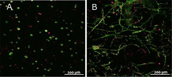
3T3 fibroblasts grown in hydrogels with PEG non cell-degradable crosslinker appeared round and in tightly packed aggregates, while cells grown in hydrogels with CD-cell degradable crossinker spread locally.

Figure 1.3T3 fibroblasts in TrueGel3D™ hydrogels made of FAST-PVA polymer modified with TrueGel3D™ RGD integrin adhesion peptide and crosslinked with either PEG non cell-degradable (left) or CD cell-degradable crosslinker (right). Pictures show collapsed stacks of confocal frames representing a height of 300 µm of the gel. Red: nuclei; green: actin cytoskeleton. Scale bar: 200 µm
Discussion
Trugel3D™ hydrogels consist of small pores at a size of only several nanometers; 3T3 fibroblasts grown in hydrogel with CD cell-degradable crosslinker spread and migrated within the gel by breaking the matrix metalloproteases (MMP) cleavable peptides included in the CD degradable crosslinker.
Fibroblasts grown in a 3D environment showed an increased level of paracrine factors1, in addition TNF receptor-1 expression and NF-kB activation2 levels were closer to in-vivo conditions. The 3D culture systems like TrueGel3D™ hydrogels present a more sensitive and biologically relevant tool for investigating various diseases involving fibroblasts.
TrueGel3D™ hydrogels are also compatible with other cell types (immune cells, cancer cell lines, epithelial cell lines, stem cells) and can be customized to study cell migration/invasion, endothelial transmigration, cell adhesion and cell differentiation.
Materials
References
To continue reading please sign in or create an account.
Don't Have An Account?