Attachment Factors for Three-dimensional Cell Culture
Section Overview
- Attachment Factors in Three-dimensional (3D) Matrices
- The Extracellular Matrix
- Extracellular Matrix Components
- ECM Attachment Factors Mediate Cell-Cell/Cell-ECM Associations
- 3D Cell Cultures
- Moving from Two- to Three-dimensional Cell Cultures
- 3D Cultures Come in Different Shapes and Sizes
- 3D Culture Composition Affects Research Application & Findings
- 3D Cultures: A Building Block in Tissue Engineering
Attachment Factors in Three-dimensional (3D) Matrices: Aspects in Tissue Engineering
Three-dimensional (3D) extracellular matrix-based cell culture systems are being used as research tools for understanding normal and disease systems, and for drug screening in vitro. The extracellular matrix (ECM) and its attachment factor components are discussed in this article in relation to their function in structural biology and their availability on the market for in vitro applications.
The Extracellular Matrix: Organizing cells into tissues
All animal cells are organized as highly complex specialized tissues. Whether the tissues are in a solid or liquid state, cells do not comprise the tissue alone, but are in constant close contact with the extracellular matrix environment. Cells secrete various types of proteins that create an intercalated mesh comprising a tissue-specialized ECM.
In general, the function of the ECM is important for:
- Tissue phenotype
- Tissue mechanical stability
- Cell motility
- Proliferation, differentiation, and morphogenesis
- Intra/Intercellular signaling
- Tissue repair
Thus, the ECM plays a key role in tissue homeostasis, in normal, pathological, and malignant states.
Extracellular matrix components: Keeping in touch
In general, the ECM is comprised of insoluble collagen fibers and soluble proteins. Different ECM components share identical structure/function modules, are interconnected, and some possess the ability to associate with cell surface components, hence their designation as attachment factors. Thus, a constant functional association exists between the ECM and tissue cells.
Descriptions of Major ECM Components:
- Collagen, a highly variable protein, is the most widespread protein in the animal kingdom. Different collagen types form rigid fibers/two-dimensional reticula, allowing solid tissues to withstand stretching.
- Proteoglycans, which have structures of a core protein attached to long polysaccharides, such as hyaluronic acid (HA), heparan sulfate, or chondroitin sulfate. This structure renders them highly hydrated creating a viscous intercellular volume, where cells can be motile during proliferation/differentiation. Some forms of these highly variable proteins span plasma membranes and are associated with intracellular signaling components, creating ECM-cytoplasm cross talk. Some forms bind growth factors and hormones, creating reservoirs and enabling presentation/ deprivation of them from tissue cells.
- Multi-adhesive matrix proteins which are intercalated and interact with collagen, cell membranes, polysaccharides, hormones and growth factors. Two major examples of these are laminin and fibronectin. Both are variable heterodimers sometimes found as a protein network, binding collagen and other ECM components, along with cell-surface integrins. Plasma fibronectin, found in the bloodstream, along with solid tissue fibronectin is associated with wound healing. Solid tissue fibronectin allows immune cell migration and plasma fibronectin participates in clotting by association with fibrin and integrins on activated platelet cell-surfaces.
It is clear from these descriptions why alterations of the ECM can result in changes in pathological states, such as enhancement of tumor cell metastatic ability. 2
ECM attachment factors mediate cell-cell/cell-ECM associations—Focal Adhesions
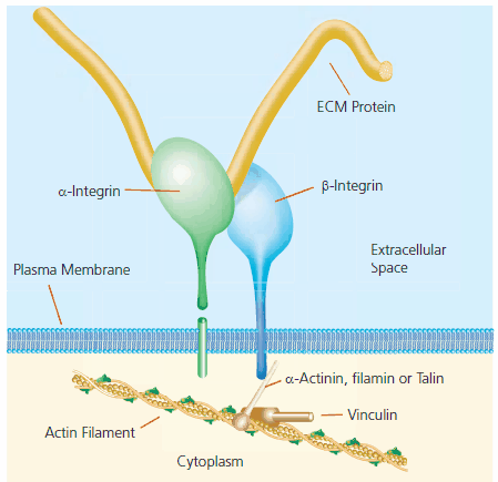
As explained previously, the structure and function of ECM components create intricate networks of cells and the ECM, leading to constant cross talk between the inner and outer cellular environment. The contact areas between the plasma membrane and the ECM are called focal adhesions. The molecular composition of these structures varies between tissues, but in general, cell-surface integrin molecules associate with both intracellular cytoskeleton-associated proteins and with ECM components (Figure 1). The ECM is a functional unit in intracellular signaling.

Figure 1. A typical focal adhesion structure.
3D Cell Cultures—“the missing link”
It has become clear that the ECM is not merely a physical scaffold for holding cells in place within tissue. The following are examples of the roles attachment factors play at the ECM-cell membrane junction in cellular function:
- Hepatocytes requiring a specialized liver-like ECM in order to express tissue-specific proteins.4,5
- The ECM acting as a reservoir of hormones and growth factors presenting or withholding specific hormones and growth factors as a result of specialized ECM-cell associations.1,6
- Bi-directional cross talk between the ECM and the cytoplasm, allowing constant cellular response to extracellular conditions. For example, the lymphocyte cell-surface molecule CD44 interacts with hyaluronic acid, enabling the initiation of the “rolling” mechanism leading to lymphocyte tissue infiltration.7
- The ECM undergoing remodeling, and cell influencing during embryonic morphogenesis and tissue repair following injury. 8
Moving from two- to three-dimensional cell cultures
Ex-vivo/in-vitro research with primary or immortalized cells is undoubtedly a convenient, relatively cheap, and reliable way to acquire preliminary information about various biological functions. However, in vitro cultures are inferior to in vivo studies in many ways. One concern is that growing cells on two-dimensional (2D) substrata creates an artificial lower and upper surface polarity, an artifact reflected in many physiological properties. However, in vivo studies are expensive, slow, and often present difficulty in isolating a single studied process/mechanism.
Three-dimensional (3D) cell cultures developed over the past years have proven to be a bridge between 2D cultures and in vivo studies, thus combining the convenience of a controlled, relatively cheap, and rapid experimental environment with physiological reliability. Researcher can now have control over both cell and matrix content, which allows mimicking of different tissues and physiological conditions, while maintaining a tissue-like structure of cells in an ECM context. Cells can be diversely manipulated prior to cultivation in the 3D matrix. The matrices relative transparency also allows visualization of processes and structures.9 For these reasons, 3D cultures have been designated “Engineered Tissues”.
3D cultures come in different shapes and sizes
In general, 3D cell cultures contain primary or immortalized cells, naïve or manipulated, seeded either on, below, or within an ECM-based matrix. Specialized cell/tissue-derived ECM matrices can then be prepared because the ECM is secreted from cells in vivo.10
Major attachment factors and ECM components are also commercially available to aid in preparing specific engineered tissues such as the following:
- ECM attachment factors for tissue culture dishes - The following attachment factors can be used either as a single matrix component or in combinations: laminin, collagen, aggrecan, fibronectin, vitronectin, and hyaluronic acid.
- ECM biomaterials - ECM gels isolated from sources like tumors (Product No. L2020 , Laminin-rich ECM and Product No. E1270, ECM gel).
- Scaffold biomaterials - Some biopolymers can be used in combination with ECM component(s) to provide 3D structure and accurate physiological function within the culture cells as shown with various tumor cells.11 (Ultra-Web® synthetic 3D in vivo-like nanofibrillar substrates from Corning®)
3D culture composition affects research application & findings
Recent advances in 3D cell culture development have led to the realization that not only are engineered tissues a valuable tool for mimicking in vivo tissue conditions, but their physical structure and attachment factor composition are critical for this behavior.
Below are examples of how 3D tissue performance is dependent on ECM and attachment factor composition:
- Engineered tumors behave differently with and without ECM additives.11 Human-derived tumors were established on 3D Poly (Lactide-co-Glycolide) (PLG) scaffolds. In the experiment, tumors were seeded on PLG alone, PLG in combination with lamininrich ECM, and on 2D cultures. Each were compared for in vivo performance in terms of proliferation, angiogenesis, hypoxia profile, and reactivity to chemotherapy. The combination of the 3D PLG structure and laminin-rich ECM yielded aggressive tumors with decreased drug-responsiveness, which best resembled tumors in vivo.
- Laminin-rich ECM gels differentiate between benign and malignant tumors.12,13 ECM gel matrix enriched with laminin, allows differentiation between benign and malignant tumors by cell morphology and proliferation. This obviously has potential use in the field of clinical diagnosis.
- Signaling pathways closely resemble in vivo networks on a specific ECM matrix.14 This work demonstrated using a basement membrane (ECM underlying epithelium)-like gel as a matrix allowing identification of signaling pathways that, when manipulated in concert, can lead to the restoration of morphologically normal breast structures or to death of highly metastatic tumor cells. This approach has potential use in designing therapeutic intervention strategies for aggressive breast cancers.
- Collagen-based 3D cultures simplify physiological genomic research . It has been shown that a 3D collagen matrix allows physiological proliferation and contraction of smooth muscle cells, accompanied by corresponding genetic expression patterns.15 3D cultures can contain genetically manipulated cells (by means of siRNA, infection, etc.) and can be relatively easy to analyze for gene expression and physiological properties. Therefore, certain 3D engineered tissues may allow for physical genetics research to:
- bypass the need for transgenic animals and extensive breeding protocols
- study gene function on a multiple genetic background.
- Both structure and composition of 3D matrices are required for functional focal adhesions.16 This work offered compelling evidence that both spatial structure and attachment factor composition of a 3D matrix influences the ability of both focal adhesions and cell biology to resemble the ones found in vivo in terms of focal adhesion-related signaling, morphology, adhesion, movement, and proliferation.
3D cultures: A building block in tissue engineering
Research has led to optimal conditions for in vitro/ex vivo work with cell cultures. The growing dilemma of complications with in vivo protocols on one hand and in vitro derived artifacts on the other hand have brought about the need for a research platform that would be reliable, resemble in vivo conditions, and allow rapid research under controlled conditions. Due to excellent academic and clinical research, and the responsiveness of the life science industry, some state-ofthe- art models exist today for growing cells in three-dimensional engineered tissues. Although some tissue types are yet to be successfully reproduced in vitro and one cannot fully mimic the complex cell types and processes intercalated in vivo, engineered 3D tissues have potential in the clinical diagnosis and treatment fields.
Related Products
References
To continue reading please sign in or create an account.
Don't Have An Account?