Protocol Guide: XTT Assay for Cell Viability and Proliferation
Introduction
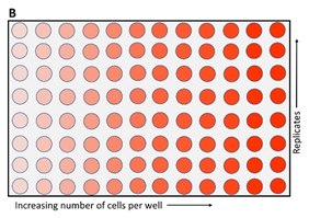
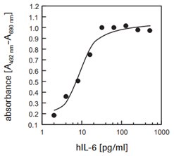
The Cell Proliferation Kit II (XTT) (Product No. 11465015001) is a colorimetric assay for the nonradioactive quantification of cellular proliferation, viability, and cytotoxicity. Sample material is either adherent or suspension cells cultured in 96-well microplates. The non-radioactive, colorimetric assay system using XTT (sodium 3´-[1- (phenylaminocarbonyl)- 3,4- tetrazolium]-bis (4-methoxy6-nitro) benzene sulfonic acid hydrate) was first described by Scudiero, P.A. et al.1, 2 and improved in subsequent years by several other investigators.3, 4 The XTT assay is used to measure cellular metabolic activity as an indicator of cell viability, proliferation and cytotoxicity. This colorimetric assay is based on the reduction of a yellow tetrazolium salt (sodium 3´-[1- (phenylaminocarbonyl)- 3,4- tetrazolium]-bis (4-methoxy6-nitro) benzene sulfonic acid hydrate or XTT) to an orange formazan dye by metabolically active cells (Figure 1).5 The formazan dye formed is soluble in aqueous solutions and is directly quantified using a scanning multiwell spectrophotometer (ELISA reader). An increase in number of living cells results in an increase in the overall activity of mitochondrial dehydrogenases in the sample. This increase directly correlates to the amount of orange formazan formed, as monitored by the absorbance (Figure 2).
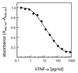
It is used for the measurement of cell proliferation in response to growth factors, cytokines and nutrients (Figure 2). It is also used for the measurement of cytotoxicity, like the quantification of tumor necrosis factor α- or β- effects (Figure 3) or the assessment of cytotoxic or growth inhibiting agents such as inhibitory antibodies.


Figure 1.Metabolism of XTT to a water-soluble orange colored formazan salt by viable cells. Schematic of an XTT assay plate.
Kit Components for The Cell Proliferation Kit II (XTT) (Product No. 11465015001)
XTT Reagent
- 5 vials containing 25 mL XTT (sodium 3´-[1- (phenylaminocarbonyl)- 3,4- tetrazolium]-bis (4-methoxy6-nitro) benzene sulfonic acid hydrate) at 1 mg/mL in RPMI 1640
Electron-coupling reagent
- 5 x 0.5 mL PMS (N-methyl dibenzopyrazine methyl sulfate)
Preparation of XTT labeling mixture
- Thaw XTT labeling reagent and electron-coupling reagent in a water bath at 37 °C. Mix each vial thoroughly to obtain a clear solution.
- To perform a cell proliferation assay (XTT) with one microplate (96 wells) mix 5 mL XTT labeling reagent with 0.1 mL electron coupling reagent.
Note: To obtain reliable results thaw and mix XTT labeling reagent and electron coupling reagent immediately before use.
Assay Protocol to Measure Cell Growth
For the determination of human interleukin-6 (hIL-6) activity on 7TD1 cells (mouse-mouse hybridoma, DSMZ, ACC 23) (Figure 2).
Additional reagents required:
- Culture medium, e.g., DMEM (Product No. D5671) containing 10% heat inactivated FBS (fetal bovine serum, Product No. 12106C), 2 mM glutamine (Product No. G6392), 0.55 mM L-arginine (Product No. A8094), 0.24 mM L-asparagine-monohydrate (Product No. A4284), 50 μM 2-mercaptoethanol (Product No. M3148), HT-media supplement (Product No. H0137) (1X), containing 0.1 mM hypoxanthine and 16 μM thymidine.
- Interleukin-6, human (hIL-6, Product No. SRP3096) (200,000 U/ml, 2 μg/mL) sterile.
- If an antibiotic is to be used, additionally supplement media with penicillin/streptomycin or gentamicin.
Protocol:
- Seed 7TD1 cells at a concentration of 4 × 10³ cells/well in 100 μL culture medium containing various amounts of IL-6 [final concentration e.g., 0.1–10 U/mL (0.001– 1 ng/mL)] into microplates (tissue culture grade, 96 wells, flat bottom).
- Incubate cell cultures for 4 days at +37°C and 5 - 6.5% CO2.
- Add 50 µL XTT labeling mixture per well and incubate for 4 h at 37 °C and 5 - 6.5% CO2.
- Measure the spectrophotometrical absorbance of the samples using a microplate (ELISA) reader. The wavelength to measure absorbance of the formazan product is between 450-500 nm according to the filters available for the ELISA reader, used. The reference wavelength should be more than 650 nm.

Figure 2.Proliferation of 7TD1 cells (mouse-mouse hybridoma) in response to recombinant human interleukin-6 (hIL-6) using XTT assay.
Assay Protocol to Measure Cytotoxicity
To determine the cytotoxic effects of human tumor necrosis factor-α (hTNF-α, T6674) on WEHI-164 cells (mouse fibrosarcoma, Product No. 87022501) (Figure 3).
Additional reagents required:
- Culture medium (e.g., RPMI 1640, Product No. R0883) containing 10% heat inactivated fetal bovine serum (FBS, Product No. 12106C), 2 mM glutamine (Product No. G6392) and 1 μg/mL actinomycin C1 (actinomycin D, Product No. A9415).
- Tumor necrosis factor-α, human (hTNF-α, Product No. T6674) (10 μg/mL), sterile.
- If an antibiotic is to be used, additionally supplement media with penicillin/streptomycin or gentamicin
Protocol:
- Preincubate WEHI-164 cells at a concentration of 1 × 10⁶ cells/ml in culture medium with 1 μg/ ml actinomycin C1 for 3 h at 37°C and 5 - 6.5% CO2.
- Seed cells at a concentration of 5 × 10⁴ cells/ well in 100 μL culture medium containing 1 μg/mL actinomycin C1 and various amounts of hTNF-α (final concentration e.g., 0.001–0.5 ng/mL) into microplates (tissue culture grade, 96 wells, flat bottom).
- Incubate cell cultures for 24 h at +37°C and 5 - 6.5% CO2.
- Add 50 μl of the XTT labeling mixture and incubate for 18 h at 37°C and 5 - 6.5% CO2.
- Measure the absorbance of the samples using a microplate (ELISA) reader. The wavelength to measure absorbance of the formazan product is between 550 and 600 nm according to the filters available for the ELISA reader, used. The reference wavelength should be more than 650 nm.

Figure 3.Determination of the cytotoxic activity of recombinant human TNF-α (hTNF-α) on WEHI-164 cells (mouse fibrosarcoma) using XTT assay
Similar Assays
MTT assay and WST-1 assay can also be used for measuring cell viability and proliferation.
Materials
References
如要继续阅读,请登录或创建帐户。
暂无帐户?