Millicell® Ultra-low Attachment Plates Knowledge Center
An important alternative to traditional 2D cell culture, 3D cell culture models closely replicate in vivo biological structures. For applications such as experimental drug discovery, cancer studies, and immunology, 3D culture models can be a more relevant avenue for researchers. Check out our 3D cell culture learning center for more information about our complete 3D cell culture portfolio.

Millicell® Ultra-low attachment plate.
Our Millicell® Ultra-low Attachment (ULA) plates are a 3D cell culture tool that promote the self-assembly of uniform spheroids. The clear, U-bottom plates allow for spheroid formation in a scaffold-free and high throughput environment. From cell seeding to fixing, staining, and imaging, Millicell® ULA plates are ideal for your cells and application. We answer your Millicell® ULA plate questions, such as:
- What are Millicell® ULA plates?
- How do they compare to other ULA plates?
- How can researchers use Millicell® ULA plates?
- Which is the ideal 3D culture plate for your application?
Explore detailed information about Millicell® ULA plates, some frequently asked questions, and some of the applications that can be performed using these plates. Request more information from our experts.
Related Products
What are Millicell® ULA plates?
Millicell® ULA cultureware are ultra-low attachment plates that promote the scaffold-free assembly of spheroids in a ready-to-use format. The plates are compatible with liquid robotic handling systems and allow researchers to form single, self-aggregated spheroid or embryoid body per well. Millicell® ULA plates support spheroid formation, assay analysis, and fluorescence imaging in the same plate.
Millicell® ULA plates are pre-coated with a stable, non-cytotoxic and non-binding surface for cells. This unique ultra-hydrophilic polymer helps researchers facilitate natural, spontaneous spheroid formation. The polymer is covalently bound to the surface of the well and has high optical clarity, making Millicell® ULA plates highly suitable for fixing, staining, and brightfield imaging and confocal microscopy in plate.

Figure 1.Schematic of spheroid formation in Millicell® ULA plates.

Figure 2.Representative brightfield imaging and confocal microscopy of A549 spheroids formed and imaged in Millicell® ULA plates.
Millicell® ULA Imaging
Millicell® ULA plates have high optical clarity and compatible with both brightfield imaging and confocal microscopy. A549 spheroids, formed in Millicell® ULA plates, were imaged in brightfield and fluorescent microscopy to demonstrate the optical clarity of the plates.
Millicell® ULA plates are recommended for confocal imaging at less than 20x objective. Confocal microscopy was performed on the ImageXpress microscope, while brightfield imaging was performed on BioTek Lionheart automated microscope.

Figure 3.Brightfield imaging of A) A549, B) HeLa, and C) MCF7 spheroid roundness with corresponding quantification.
Evaluating Spheroid Formation and Roundness in Millicell® ULA Plates
Millicell® ULA plates help facilitate uniform spheroid formation for scalable cell culture applications. We used the plates to quantitatively evaluate spheroid formation through calculations of spheroid roundness and circularity.
We provide a spheroid formation protocol using A549, HeLa, and MCF7 cells and demonstrate how to calculate roundness and circularity, two quantitative measurements that demonstrate spheroid homogeneity and compactness. By quantifying spheroid roundness and circularity, researchers can also monitor the extracellular matrix that formed around the spheroid.

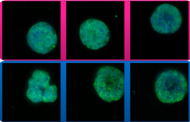
Figure 4.Confocal microscopy of HeLa cells using a 10X objective. Top: HeLa spheroids formed in Millicell® ULA plates; Bottom: HeLa spheroids formed in Competitor A ULA plates.
Millicell® ULA Plates vs Competition ULA Plates
ULA plates, or ultra-low attachment plates, contain a hydrogel layer that is covalently bound to the surface of the well. This hydrogel layer inhibits cell attachment to the plate, minimizing protein absorption and enzyme and cell activation and helping to form spheroids in the wells.
We compared our Millicell® ULA plates to a competitor ULA plate (Competitor A), to demonstrate how spheroid formation, roundness, and circularity can vary between the two plates. Ergonomically, there are subtle differences between the plate and lid shapes in the two plates, and Millicell® ULA plates have clearer labeling on the surface of the plate. We monitored the spheroid formation of several cell lines, whether the spheroid lines grew successfully, and if there was a difference in plate performance.
Related Resources
- Corning® Spheroid Microplates
Understand how Corning® Spheroid Microplates can be used for generating and culturing tumor spheroids in a 96-well spheroid microplate format.
- Millicell® Ultra-Low Attachment (ULA) Plate FAQs
Learn how to grow spheroid cultures in ULA plates, the common applications and benefits of ULA plates for 3D cell culture, Millicell® ULA plate dimensions, plate handling, troubleshooting, and more.
如要继续阅读,请登录或创建帐户。
暂无帐户?