HPA018425
Anti-G3BP2 antibody produced in rabbit

Prestige Antibodies® Powered by Atlas Antibodies, affinity isolated antibody, buffered aqueous glycerol solution
别名:
Anti-G3BP-2, Anti-GAP SH3 domain-binding protein 2, Anti-Ras GTPase-activating protein-binding protein 2
登录查看公司和协议定价
所有图片(9)
About This Item
推荐产品
生物来源
rabbit
质量水平
偶联物
unconjugated
抗体形式
affinity isolated antibody
抗体产品类型
primary antibodies
克隆
polyclonal
产品线
Prestige Antibodies® Powered by Atlas Antibodies
表单
buffered aqueous glycerol solution
种属反应性
human
增强验证
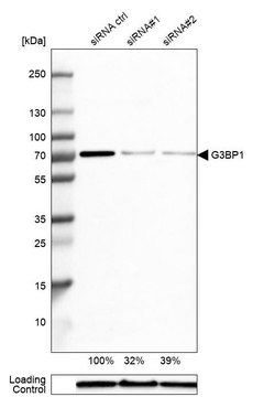
orthogonal RNAseq
independent
Learn more about Antibody Enhanced Validation
技术
immunohistochemistry: 1:50-1:200
免疫原序列
KNLEELEEKSTTPPPAEPVSLPQEPPKPRVEAKPEVQSQPPRVREQRPRERPGFPPRGPRPGRGDMEQNDS
UniProt登记号
运输
wet ice
储存温度
−20°C
靶向翻译后修饰
unmodified
基因信息
human ... G3BP2(9908)
一般描述
The gene G3BP2 (GAP SH3 domain-binding protein-2) has been mapped to human chromosome 4q21.1. G3BP2 is ubiquitously expressed and is mainly present in the cytoplasm. However, it is capable of shuttling into the nucleus in cell-cycle dependent manner. G3BP2 include an NTF2 (nuclear transport factor)-like domain and two RNA-binding motifs.
免疫原
Ras GTPase-activating protein-binding protein 2 recombinant protein epitope signature tag (PrEST)
应用
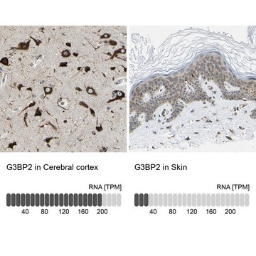
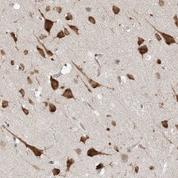
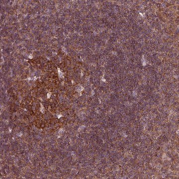
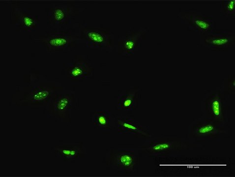
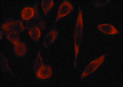
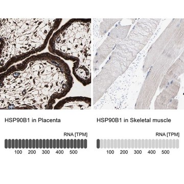
Anti-G3BP2 antibody produced in rabbit, a Prestige Antibody, is developed and validated by the Human Protein Atlas (HPA) project . Each antibody is tested by immunohistochemistry against hundreds of normal and disease tissues. These images can be viewed on the Human Protein Atlas (HPA) site by clicking on the Image Gallery link. The antibodies are also tested using immunofluorescence and western blotting. To view these protocols and other useful information about Prestige Antibodies and the HPA, visit sigma.com/prestige.
生化/生理作用
GAP SH3 domain-binding protein-2 (G3BP2) is identified as a gene for predicting the presence of lymph node metastasis in primary oral squamous cell carcinoma. G3BP2 is a negative modulator of p53 function. Depletion of G3BP2 leads to up-regulation of p53 and its overexpression leads to the cytoplasmic redistribution of p53. G3BP2 binds mechanomediator TWIST1 in the cytoplasm. Loss of G3BP2 leads to nuclear localization of TWIST1 which in response to biomechanical signals induce epithelial-mesenchymal transition, promoting tumor invasion and metastasis. Overexpression of G3BP2 induces formation of cytoplasmic RNA granules called stress granules (SGs). Knockdown of G3BP2 reduces the number of SG-positive cells. During Semliki Forest virus infection, the C-terminal domain of the viral nonstructural protein-3 forms a complex with G3BP2 and inhibits the formation of SGs on viral mRNAs, thus impairing antiviral defense.
特点和优势
Prestige Antibodies® are highly characterized and extensively validated antibodies with the added benefit of all available characterization data for each target being accessible via the Human Protein Atlas portal linked just below the product name at the top of this page. The uniqueness and low cross-reactivity of the Prestige Antibodies® to other proteins are due to a thorough selection of antigen regions, affinity purification, and stringent selection. Prestige antigen controls are available for every corresponding Prestige Antibody and can be found in the linkage section.
Every Prestige Antibody is tested in the following ways:
Every Prestige Antibody is tested in the following ways:
- IHC tissue array of 44 normal human tissues and 20 of the most common cancer type tissues.
- Protein array of 364 human recombinant protein fragments.
联系
Corresponding Antigen APREST73970
外形
Solution in phosphate-buffered saline, pH 7.2, containing 40% glycerol and 0.02% sodium azide
法律信息
Prestige Antibodies is a registered trademark of Merck KGaA, Darmstadt, Germany
免责声明
Unless otherwise stated in our catalog or other company documentation accompanying the product(s), our products are intended for research use only and are not to be used for any other purpose, which includes but is not limited to, unauthorized commercial uses, in vitro diagnostic uses, ex vivo or in vivo therapeutic uses or any type of consumption or application to humans or animals.
未找到合适的产品?
试试我们的产品选型工具.
储存分类代码
10 - Combustible liquids
WGK
WGK 1
闪点(°F)
Not applicable
闪点(°C)
Not applicable
法规信息
常规特殊物品
Weina Wang et al.
Advanced science (Weinheim, Baden-Wurttemberg, Germany), 11(6), e2305068-e2305068 (2023-12-13)
Primary cilia are conserved organelles in most mammalian cells, acting as "antennae" to sense external signals. Maintaining a physiological cilium length is required for cilium function. MicroRNAs (miRNAs) are potent gene expression regulators, and aberrant miRNA expression is closely associated
Ying Sun et al.
Oncogene, 38(24), 4856-4874 (2019-02-26)
Identification of molecular alterations driving breast cancer progression is critical for the development of effective therapy. In this study, we show that the level of α-parvin is elevated in triple-negative breast cancer cells. The depletion of α-parvin from triple-negative breast
M M Kim et al.
Oncogene, 26(29), 4209-4215 (2007-02-14)
Inactivation of the p53 tumor suppressor pathway is a critical step in human tumorigenesis. In addition to mutations, p53 can be functionally silenced through its increased degradation, inhibition of its transcriptional activity and/or its inappropriate subcellular localization. Using a proteomic
Fátima Solange Pasini et al.
Acta oncologica (Stockholm, Sweden), 51(1), 77-85 (2011-10-12)
Previous knowledge of cervical lymph node compromise may be crucial to choose the best treatment strategy in oral squamous cell carcinoma (OSCC). Here we propose a set four genes, whose mRNA expression in the primary tumor predicts nodal status in
Marc D Panas et al.
Molecular biology of the cell, 23(24), 4701-4712 (2012-10-23)
Dynamic, mRNA-containing stress granules (SGs) form in the cytoplasm of cells under environmental stresses, including viral infection. Many viruses appear to employ mechanisms to disrupt the formation of SGs on their mRNAs, suggesting that they represent a cellular defense against
我们的科学家团队拥有各种研究领域经验,包括生命科学、材料科学、化学合成、色谱、分析及许多其他领域.
联系技术服务部门