feature
according to Highman
IVD
for in vitro diagnostic use
dilution
(for human-medical cell diagnosis)
application(s)
clinical testing
diagnostic assay manufacturing
hematology
histology, detection
storage temp.
15-25°C
Quality Level
General description
The Congo Red staining kit - Kit for the detection of amyloid acc. to Highman, is used for human-medical cell diagnosis and serves the purpose of the histological investigation of sample material of human origin for example histological sections of e. g. the kidney, the intestine, or the liver.
This Congo red staining kit contains all the reagents necessary for staining amyloid in histological tissues. Amyloid is a homogenous structure made up of protein fibrils (each between 8 and 15 nm in diameter) that can be stained eosinophilically, which e. g. in the case of amyloidosis forms deposits in the intercellular space. All deposits of amyloid contain similar protein fibrils that are resistant to the body′s natural defence mechanisms and that once they have formed cannot be eliminated.The Congo red staining principle is based on the formation of hydrogen bridge bonds with the carbohydrate component of the substrate. Congo red is an anionic dye and is capable of depositing itself in amyloid fibrils, which then exhibit a conspicuous dichroism under polarized light. The tissue stained with Congo red appears orange-red under the transmitted-light microscope; under polarized light, however, the amyloid deposits show up as brilliant green double-refraction images against a dark background. Other structures also stained by Congo red, e. g. collagen, however are not visualized under polarized light. Staining may be technically difficult when the paraffin sections used are too thin (<5 μm) or when the tissue is too strongly over-stained.
The kit is sufficient for up to 50 applications. This product is registered as IVD and CE marked. For more details, please see instructions for use (IFU). The IFU can be downloaded from this webpage.
Application
Kit for the detection of amyloid acc. to Highman
Analysis Note
Suitability for microscopypasses testAmyloidpink to red; in polarized ligt green metachromasisNucleidark blueConnective tissuelight red
signalword
Danger
hcodes
Hazard Classifications
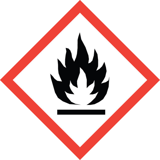
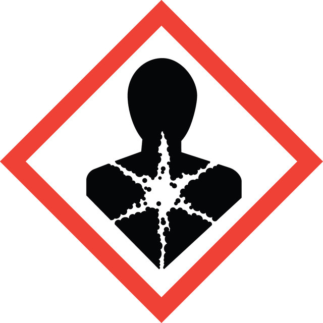
Carc. 1B - Eye Irrit. 2 - Flam. Liq. 2 - Met. Corr. 1 - Skin Irrit. 2
存储类别
3 - Flammable liquids
法规信息
监管及禁止进口产品
此项目有
我们的科学家团队拥有各种研究领域经验,包括生命科学、材料科学、化学合成、色谱、分析及许多其他领域.
联系客户支持

