3D Cell Culture Workflow Tools
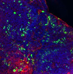
Figure 1. Characterization of human colon organoids using immunocytochemical stains for Mucin-1, F-actin, and DAPI.
What is 3D Cell Culture?
Cells in their natural environment have constant interactions with their extracellular matrix (ECM) and other cells, regulated by complex biological functions. Three-dimensional (3D) cell culture systems allow cells to grow and interact with their surroundings in all three dimensions.
By utilizing 3D cell culture models, researchers can simulate the in vivo physiological microenvironment. These models also provide better predictive in vitro cell models for applications including cancer research, drug discovery, and regenerative medicine. Transitioning to 3D cell culture can bring together the biological relevance of animal models with the accessibility of traditional 2D models.
3D Culture Models
We offer an extensive portfolio of tools, protocols, and technologies to support 3D cell culture applications such as drug permeability, ADME/toxicity assays, cancer, and autoimmune disorders.
Explore our Knowledge Center below and discover answers to questions about organoids, hydrogels, and organotypic cultures. Request more information here.
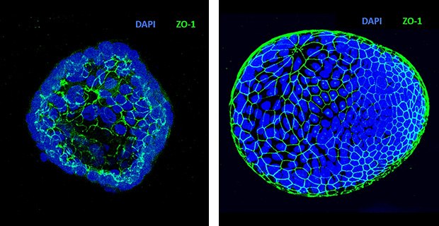
Figure 2. Apical-out Human Colon Organoids Generation. The epithelial cell polarity was reversed using the apical-out organoid culture protocol and 3dGRO™ Human Colon Intestinal Organoids. Polarity reversion was determined using DAPI (blue) and ZO-1 (MABT339, Green) immunocytochemical staining.
Organoids are mini-organs derived from stem cells and grown in near-native culture conditions that allow for the development of functional 3D structures preserving key physiological function of the original tissue. They can be derived from patient tissues (patient-derived organoids; PDOs) or induced pluripotent stem cells (iPSCs).
Organoids have been used to study organ-specific disorders and diseases including related cancers, inflammatory bowel disease, and cystic fibrosis. They are also used to study viral respiratory disease, drug transport, drug permeability, ADME/Tox, and organogenesis. Our portfolio includes organoids derived from patient tissues and induced pluripotent stem cells; we also provide expert answers to frequently asked questions about organoid culture.
We have now expanded our organoid portfolio to include HUB Organoids. The founder of adult stem cell-derived organoid technology, HUB Organoids specializes in the application of Organoid Technology to preclinical testing, drug discovery, and therapeutic development. Our healthy and diseased patient-derived organoid biobanks include gastrointestinal, lung cancer, and pancreatic ductal adenocarcinoma organoids, and are subjected to rigorous quality control protocols. The biobanks are stable for long-term culture and cryopreservation.
We have also rounded out our portfolio to include access to HUB Organoid screening services. Accelerate the discovery of therapeutic treatments, such as for infectious diseases, oncology, and toxicology investigations, and closely mimic human biology with this novel platform.
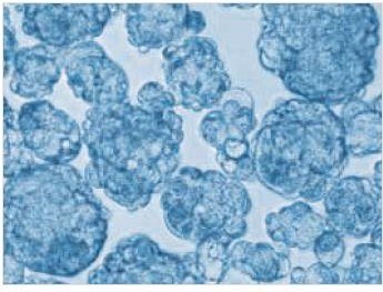
Figure 3. MCF-7 breast cancer cell culture. Tumorsphere formation using MCF-7 cell lines after 10 days of serial passage.
Formed by the self-aggregation of cells, spheroid cultures are excellent models of tumor-related disorders and diseases such as cancer. Because spheroids have surface cells and buried cells, these models are commonly used to tumor formation, tumor hypoxia, and drug penetration.
Spheroids are easily amenable to both high throughput and co-culture applications. They can be grown using scaffold-free technologies such as ultra-low attachment (ULA) plates or scaffold-based technologies such as hydrogels.
Hydrogels are a cellular tool that researchers use to mimic the natural cellular environment in vitro. As a water-swollen network of polymers, hydrogels remain in the liquid state at 4˚C. This allows researchers to ebbed cells before the gel forms at incubation temperatures of 37˚C. The cells then become encapsulated in the gel and can be cultured in an environment that is physiologically relevant.
Need to choose a hydrogel? Learn about the key considerations in hydrogel selection for 3D cell culture applications, including:
- Natural vs. synthetic hydrogels
- Biological relevance, reproducibility, reactivity, and ease of use
- Customization, with formats including powders, liquids, kits, and pre-cast microplates
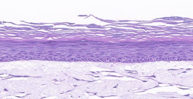
Figure 4. In vitro organotypic skin culture model. H&E staining showing epidermal and dermal stratified layers using an in vitro organotypic model.
Organotypic culture systems (OCSs) culture organs in vitro that were collected from an organism. These models preserve normal organ architecture and interactions while supporting the formation of nearly normal stratified epidermis cells. They can be used to mimic complex pathologies of the human skin, airway, intestine, liver, heart, and neurological tissues.
Organotypic models are commonly used to study a variety of related cancers, aging, autoimmune disorders, infection, tissue microenvironment, and tissue development. These systems are typically cultured using scaffold-based technologies such as porous membrane-based cell culture inserts.
Figure 5. Time-lapse video of Forskolin Induced Swelling assay on 3dGRO® Human Colon Organoids at 20 µM Forskolin after 4.3 hours.
Culturing organoids, spheroids, and other 3D models often require specific tools to support the establishment of the cultures. Researchers could use separate scaffolds for their cultures, or they can culture cells directly into 3D cell culture plates. Plates with ultra-low attachment (ULA) coatings, or that contain additional microwells promote organoid, tumoroid, and spheroid formation in a controlled 3D environment.
Millicell® Ultra-low Attachment (ULA) Plates
Millicell®ULA plates promote scaffold free self-assembly of spheroids using a stable, non-binding polymer. The plates contain a ultra-hydrophilic polymer that facilitates spontaneous and natural spheroid formation. Millicell®ULA plates have high optical clarity, making them suitable for both brightfield imaging and confocal microscopy.
We cultured multiple spheroid forming cells using Millicell® ULA plates. Using the plates, we developed quantitative measurements of spheroid formation by calculating spheroid circularity and roundness.
Millicell® Microwell Plates
Millicell® Microwell plates contain an array of ultra-dense, U-bottom microwells (W) and hydrogel. The addition of microwells promote reproducible organoid culture for high throughput and screening applications. The microwells also allow researchers to generate up to hundreds of homogeneous organoids in a single well. As the organoids form in a single focal plane, Millicell® Microwell plates support organoid seeding, imaging, and analysis in the plate.
Millicell® Microwell plates are ideal for multiple applications, including blood brain barrier penetration assays and T cell killing assays.
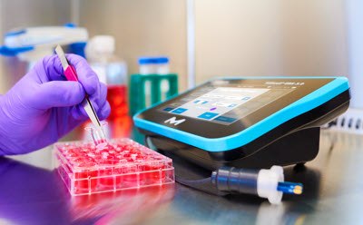
When researchers are looking for more biologically relevant methods than 2D cell culture, they often turn to 2.5D culture methods. In 2.5D cell culture, researchers grow cells on coated surfaces such as a biological coating or synthetic polymer, enhancing cell growth, attachment, and differentiation. Researchers can move from 2D to 2.5D culture applications with our collection of tools, including Millicell® inserts and the Millicell® ERS 3.0 Digital Voltohmmeter.
Millicell® ERS 3.0 Digital Voltohmmeter
The Millicell® ERS 3.0 Digital Voltohmmeter acquires highly stable TEER measurements, automatically saves your data, and includes an easy-to-use touchscreen interface, a standing in-well probe, a plug-in or battery pack power source, and an adjustable electrode that is compatible with a wide variety of cell culture inserts, including Millicell® cell culture inserts.
TEER data from the Millicell® ERS 3.0 Digital Voltohmmeter can easily be exported via USB drive, ethernet, or uploaded to the cloud. Our Millicell® Cloud features comprehensive measurement view, enabling visualization and analysis your data in various formats.
Millicell® Cell Culture Inserts
TEER readers are often used to monitor and measure epithelial monolayer barrier formation in 2.5D experiments. Researchers perform barrier formation assays on hanging inserts with porous membranes, with cells forming confluent monolayers when there is an increase in resistance measurements. Millicell® cell culture inserts allow researchers to study the apical and basal surfaces of the monolayer and are ideal for co-culture applications.
Materials
Moving from 2D to 3D
3D cell culture is an in vitro method for researchers who want to more closely model physiological conditions. It is often more relevant than using traditional 2D methods. 3D cell culture methods promote interactions with other cells and with the physical environment. These interactions can regulate multiple cellular functions, including transcription regulation and apoptosis.
Scaffold-free technologies
One method of 3D cell culture is the scaffold-free model. Scaffold-free 3D cell culture techniques allow cells to self-assemble and form non-adherent cell aggregates, clusters, spheroids, or tumorspheres.
Scaffold-based technologies
In scaffold-based 3D cell culture methods, 3D scaffolds provide structural support for cell attachment and tissue development. This recapitulates elements of the native extracellular matrix (ECM) or cellular environment.
For researchers studying drug safety and efficacy, ADME/toxicity, and biotherapeutics development, 3D cell culture models can reduce the time spent in setting up and performing assays.
Related Content
- T-Cell Migration Assays Using Millicell® Cell Culture Inserts
Learn how to perform cell migration assays in vitro using Millicell® hanging cell culture inserts and the suspension T-cell lines Jurkat and primary CD4+ cells. Monitor migration by flow cytometry and EZ-MTT assays.
- Tumor Spheroid Formation Assay
3D cell culture protocol: the tumor spheroid formation assay using a serum-free and xeno-free cancer stem cell culture media.
- 3D Bioprinting: Bioink Selection Guide
Bioinks can be 3D bioprinted into functional tissue constructs for drug screening, disease modeling, and in vitro transplantation. Choose the Bioinks and method for specific tissues engineering applications.
- Webinar: 3D Organ-on-a-Chip Applications Using the AIM Biotech Chip
3D Organ-on-a-Chip Applications Using the AIM Biotech Chip
- Attachment Factors for 3-Dimensional Cell Culture
The extracellular matrix (ECM) and its attachment factor components are discussed in this article in relation to their function in structural biology and their availability for in vitro applications.
- Cellular Fluorescence Imaging with Millicell® Cell Culture Inserts
Millicell® hanging cell culture inserts for staining and microscopic analysis directly from the insert. Learn to utilize hanging cell culture inserts in barrier measurements.
- /deepweb/assets/sigmaaldrich/marketing/global/documents/425/972/3dcc-br6480en-ms.pdf
Related Product Resources
- Tumor Spheroid Formation Assay
3D cell culture protocol: the tumor spheroid formation assay using a serum-free and xeno-free cancer stem cell culture media.
- Bioink Selection for 3D Bioprinting
Bioinks enable 3D bioprinting of tissue constructs for drug screening and transplantation; select suitable bioinks for specific tissue engineering.
- Cell Imaging Instruments
Enhance cell analysis: Utilize imaging tools to assess, analyze cell states. Affordable solutions for live, digital imaging, expanding lab capabilities.
- Webinar: 3D Organ-on-a-Chip Applications Using the AIM Biotech Chip
3D Organ-on-a-Chip Applications Using the AIM Biotech Chip
- Scaffolds for 3D Cell Culture
Cells grown on structural scaffolds engineered to mimic the ECM can be powerful, predictive models of physiological processes. Our wide range of advanced 3D cell culture scaffolds includes nanofiber, collagen, polystyrene, and polycaprolactone materials.
- Attachment Factors for Three-dimensional Cell Culture
The extracellular matrix (ECM) and its attachment factor components are discussed in this article in relation to their function in structural biology and their availability for in vitro applications.
- Hydrogels
Cells embedded in 3D hydrogels can polarize, differentiate and have functional attributes that more closely resemble physiological tissues. Choose naturally derived hydrogels or reconstitute a hydrogel from synthetic materials to develop predictive cellular models.
- /deepweb/assets/sigmaaldrich/marketing/global/documents/425/972/3dcc-br6480en-ms.pdf
Cultivate reality with state-of-the-art 3D cell technologies including tumor spheroids, stem cell organoids, patient-derived organoids, and tissue engineering via 3D bioprinting.
如要继续阅读,请登录或创建帐户。
暂无帐户?