The Power of High Sensitivity Immunoassays In Immunology Research
Immunology research encompasses many types of studies, including immuno-oncology, inflammation, autoimmunity, and viral research areas. Immunoassays are vital to this research to detect biomarkers such as interferons, interleukins, immunoglobulins, and more. Learn how high sensitivity immunology assays, like SMC® high sensitivity immunoassay kits, can be used to enhance the power of your immunology studies.
Section Overview
Why Is Sensitivity Important in Immunoassays for Immunology Research?
Immunoassays play a critical role in researching the immune system because they help analyze biomarkers like interferons, interleukins, immunoglobulins, and other cytokines/chemokines. The sensitivity of immunoassays is important in immunology research because higher sensitivity allows researchers to thoroughly characterize biomarkers of interest. With this capability, they can dive deeper into immune pathways and gain a better understanding of immune mechanisms.
SMC® High Sensitivity Immunology Assays
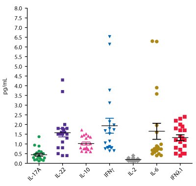
Harness the power of SMC® high sensitivity immunology immunoassay kits to analyze immune biomarkers on a deeper level. These assays can be combined with sub-picogram sensitivity and a user-friendly experience on the SMCxPRO® ultrasensitive immunoassay platform to detect low-level biomarkers. See how SMC® kits can be used in immunology research with the example data below (Figure 1).

Figure 1.Apparently healthy human serum and EDTA plasma samples were analyzed using SMC® immunoassays for IL-17A, IL-22, IL-10, IFNγ, IL-2, IL-6, and IFNλ1 on the SMCxPRO® platform. Sample concentrations are shown in pg/mL.
Th1/Th2 Cytokines
The Th1 and Th2 subsets of T helper cells function in allergy and autoimmune diseases. Th1 cells secrete cytokines, including IFNɣ, to protect against viral and bacterial infection.1 Th2 cells secrete IL-4, IL-5, IL-10, and IL-13 to activate B cells and antibody production. We offer SMC® kits to detect Th1/Th2 cytokines with LLOQ as low as 0.03 pg/mL.
Th17 Cytokines
Th17 cells emerged as a new CD4+ T helper subset, secreting IL-17A, IL-17F, IL-22, and other cytokines that play a role in tissue inflammation and function to protect against extracellular pathogens.2 The Th17 subset has been found to play a significant role in immune-mediated diseases including rheumatoid arthritis, psoriasis, and systemic lupus erythematosus.3 Other disease associations include multiple sclerosis, atopic dermatitis, and asthma. Measure sub-picogram levels of key Th17 targets with ultrasensitive SMC® immunology assays.
Other Cytokines, Chemokines, and Growth Factors
Cytokines, chemokines, and growth factors play an important role in immune system functions and have autocrine, paracrine, and endocrine signaling mechanisms. Their pleiotropic immunomodulatory properties give them the ability to respond against various stimuli through immune response regulation, either by stimulating or preventing inflammation. Growing research continues to highlight the role of the immune system in every facet of human health and disease.
Immuno-Oncology Checkpoint Proteins
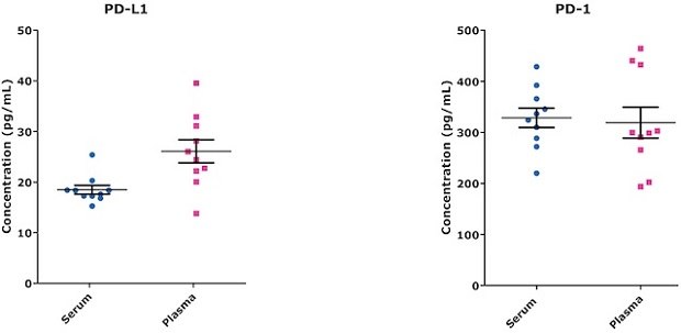
Immuno-oncology has significantly transformed cancer treatment demonstrating the achievement of remission in patients. This breakthrough has been made possible by the use of immune checkpoint inhibitors and cellular therapies. Despite these achievements, challenges remain for certain types of cancer. To overcome these challenges, researchers are focusing on the identification and validation of secreted biomarkers. These biomarkers, found in serum or plasma, play a crucial role in identifying patients who are likely to respond well to specific therapeutic interventions. Key checkpoint targets, such as PD-L1 and PD-L2, have emerged as valuable prognostic indicators in specific cancer types. Additionally, the elevated secretion of interferon gamma in response to cellular therapy serves as an early indication of cytokine release syndrome (CRS), which is an adverse response that needs to be carefully monitored and managed. Highly sensitive immunoassays are crucial for this research as they enable the accurate detection and quantification of low levels of biomarkers in samples. This level of sensitivity is essential for research aiming to identify subtle changes in biomarker expression and ensure reliable and precise patient stratification, ultimately leading to the enhancement of immuno-oncology therapy effectiveness. Figure 2 provides example endogenous sample concentrations of PD-L1 and PD-1 detected using ultrasensitive SMC® immunoassays.

Figure 2.Endogenous sample concentrations using the SMC® Human PD-1 and PD-L1 High Sensitivity kits. Healthy donor matched serum (n=10) and EDTA plasma (n=10) were used.
Related Webinars
Watch the webinars below to see case studies of SMC® technology being used in immunology research.
- A Sensitive Method to Quantify HIV-1 Antibody in Mucosal Samples (can be viewed only in the US & Canada)
- Development of Ultrasensitive Immunoassays for SARS-CoV-2 Antibody Detection
For Research Use Only. Not For Use In Diagnostic Procedures.
Related Products
References
如要继续阅读,请登录或创建帐户。
暂无帐户?