跳转至内容
Merck
CN

葡聚糖

货号:9004-54-0

结构

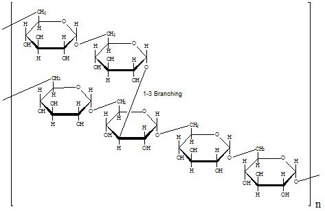
葡聚糖是葡萄糖酐聚合物。  它由大约 95% 的alpha-D-(1-6)键合组成。其余的 a (1-3) 键合解释了葡聚糖的分支。1,2,3 分支长度的冲突数据意味着平均分支长度小于三个葡萄糖单位。4,5 但是,其他方法表明存在大于 50 个葡萄糖单位的分支。6,7

已发现天然葡聚糖的分子量(MW)在900万至5亿之间。8,9,10 较低MW的葡聚糖表现出较低的分支化4,并且具有更窄的MW分布范围。11 MW大于10,000的葡聚糖表现高度支化。  随着MW的提高,葡聚糖分子会具有更好的对称性。7,12,13 MW在2,000至10,000之间的葡聚糖分子可展现出可膨胀线圈的特性。1212 MW小于2,000的葡聚糖更多是棒状的。14

葡聚糖的分子量通过以下一种或多种方法测定:低角度激光散射15、尺寸排阻色谱法16、铜络合17和蒽酮试剂18、比色法还原糖测定和粘度12

比旋度:[α]=+199° 11

比旋度

图 1.比旋度

产品信息

我们提供来自肠膜明串珠菌菌株B 512的葡聚糖。  通过有限的水解和分馏生成各种分子量。我们供应商采用的确切方法已申请专利。分馏可通过尺寸排阻色谱16 或乙醇分级,其中最大分子量的葡聚糖首先沉淀。19

制备说明

右旋糖酐具有很好的水溶性,最高分子量的右旋糖酐除外,货号D5501(MW范围 = 500万至4000万)。  我们测试葡聚糖在水中的浓度通常超过30 mg/mL的溶解度。
葡聚糖也可自由溶于DMSO、甲酰胺、乙二醇和甘油。11
葡聚糖中性水溶液可通过在110-115 °C下高压灭菌30至45分钟而进行灭菌。11葡聚糖可在高温下被强酸水解。葡聚糖的末端还原基团可在碱性溶液中氧化。11

操作流程

右旋糖酐是一种高分子量、惰性、水溶性聚合物,已被广泛应用于生物医学领域。

未修饰的葡聚糖的应用:

血浆填充剂

右旋糖酐溶液已被用作血浆填充剂。20

10% 葡聚糖溶液 (分子量40,000) 比血浆蛋白产生略高的胶体渗透压。

据悉,0.9% 氯化钠或 5% 葡萄糖中的 10% 右旋糖酐 (分子量40,000) 溶液可用作术后血栓栓塞性疾病的短期血浆增量剂。  输注后,24小时后尿液中约70%的葡聚糖(分子量40,000)不变。粪便中有少量排出。  剩余的右旋糖酐缓慢代谢为葡萄糖。

6% 右旋糖酐溶液 (分子量70,000) 的胶体渗透压与血浆蛋白相似。分子量大于 50,000 的右旋糖酐倾向于缓慢扩散穿过毛细血管壁,并缓慢代谢为葡萄糖。大约50%输注的右旋糖酐(分子量70,000)在24 小时内尿中排泄不变20

离心/细胞和细胞器分离

含右旋糖酐 (分子量250,000) 的胶体溶液已用于离心分离血液中聚集的血小板21 白细胞22 和淋巴细胞23

葡聚糖 (分子量40,000) 已被用于分离完整的细胞核。24

蛋白质沉淀

在免疫扩散应用中,葡聚糖已被用于增强抗体-抗原复合物的沉淀和敏感性。

将右旋糖酐 (分子量80,000) 以最大80 mg/mL注入免疫电泳凝胶中。25

右旋糖酐(分子量 250,000 和 2,000,000)也用于类似的应用。26

抑制血小板聚集

较低浓度的右旋糖酐 (分子量 10,000-40,000) 已用于抑制血小板聚集。27

参考文献

1.
Rankin JC, Jeanes A. 1954. Evaluation of the Periodate Oxidation Method for Structural Analysis of Dextrans. J. Am. Chem. Soc.. 76(17):4435-4441. https://doi.org/10.1021/ja01646a046
2.
Dimler RJ, Wolff IA, Sloan JW, Rist CE. 1955. Interpretation of Periodate Oxidation Data on Degraded Dextran. J. Am. Chem. Soc.. 77(24):6568-6573. https://doi.org/10.1021/ja01629a044
3.
Van Cleve JW, Schaefer WC, Rist CE. 1956. The Structure of NRRL B-512 Dextran. Methylation Studies2. J. Am. Chem. Soc.. 78(17):4435-4438. https://doi.org/10.1021/ja01598a064
4.
Lindberg B, Svensson S, Sjövall J, Zaidi NA. 1968. Structural Studies on Dextran from Leuconostoc mesenteroides NRRL B-512.. Acta Chem. Scand.. 221907-1912. https://doi.org/10.3891/acta.chem.scand.22-1907
5.
Larm O, Lindberg B, Svensson S. 1971. Studies on the length of the side chains of the dextran elaborated by Leuconostoc mesenteroides NRRL B-512. Carbohydrate Research. 20(1):39-48. https://doi.org/10.1016/s0008-6215(00)84947-2
6.
Bovey FA. 1959. Enzymatic polymerization. I. Molecular weight and branching during the formation of dextran. J. Polym. Sci.. 35(128):167-182. https://doi.org/10.1002/pol.1959.1203512813
7.
Senti FR, Hellman NN, Ludwig NH, Babcock GE, Tobin R, Glass CA, Lamberts BL. 1955. Viscosity, sedimentation, and light-scattering properties of fraction of an acid-hydrolyzed dextran. J. Polym. Sci.. 17(86):527-546. https://doi.org/10.1002/pol.1955.120178605
8.
Arond LH, Frank HP. 1954. Molecular Weight Distribution and Molecular Size of a Native Dextran. J. Phys. Chem.. 58(11):953-957. https://doi.org/10.1021/j150521a006
9.
Elias VH. 1959. Ultrazentrifugen- und Diffusionsmessungen an nicht-Newtonschen Lösungen nativer Dextrane. Über extrem große Makromoleküle. IV. Makromol. Chem.. 33(1):166-180. https://doi.org/10.1002/macp.1959.020330112
10.
Antonini E, Bellelli L, Bruzzesi MR, Caputo A, Chiancone E, Rossi-Fanelli A. 1964. Studies on dextran and dextran derivatives. I. Properties of native dextran in different solvents. Biopolymers. 2(1):27-34. https://doi.org/10.1002/bip.1964.360020105
11.
Supplier's data..
12.
Granath KA. 1958. Solution properties of branched dextrans. Journal of Colloid Science. 13(4):308-328. https://doi.org/10.1016/0095-8522(58)90041-2
13.
Wales M. 1979. J.Polym. Sci. 66101.
14.
GEKKO K. 1981. Solution Properties of Dextran and Its Ionic Derivatives.415-438. https://doi.org/10.1021/bk-1981-0150.ch029
15.
Gaylord NG, Beaman RG. 1960. Techniques of polymer characterization. P. W. Allen. Academic Press, New York; Butter-worths, London, 1959. xiv + 256 pp. $9.50.. J. Appl. Polym. Sci.. 3(7):128-128. https://doi.org/10.1002/app.1960.070030723
16.
Granath KA, Flodin P. 1961. Makromol. Chem.. 48(1):160-171. https://doi.org/10.1002/macp.1961.020480116
17.
1953. Journal of Research of the National Bureau of Standards. 5081.
18.
Jermyn M. 1975. Increasing the sensitivity of the anthrone method for carbohydrate. Analytical Biochemistry. 68(1):332-335. https://doi.org/10.1016/0003-2697(75)90713-7
19.
Cramér H. 1949. On the factorization of certain probability distributions. Ark. Mat.. 1(1):61-65. https://doi.org/10.1007/bf02590468
20.
LIPMAN AG. 1993. MARTINDALE: ?Martindale - the Extra Pharmacopoeia? (30th ed), edited by J. E. F. Reynolds. 2(2):124-124. https://doi.org/10.1111/j.2042-7174.1993.tb00740.x
21.
Minor A, Burnett L, State J N. 1953. 53547.
22.
Boyum A, Scan J. 1968. Clin. Lab. Invest.. 21(97):31.
23.
Harris R, Ukaejiofo EO. 1970. Tissue Typing Using a Routine One-Step Lymphocyte Separation Procedure. Br J Haematol. 18(2):229-236. https://doi.org/10.1111/j.1365-2141.1970.tb01436.x
24.
HONDA SI, HONGLADAROM T, LATIES GG. 1966. A New Isolation Medium for Plant Organelles. J Exp Bot. 17(3):460-472. https://doi.org/10.1093/jxb/17.3.460
25.
Hellsing K. 1969. Immune reactions in polysaccharide media. The composition of the antigen?antibody complexes in the precipitin reaction. 114(1):141-144. https://doi.org/10.1042/bj1140141
26.
Ceska M. 1969. Human growth hormone-anti-(growth hormone) immunoprecipitates in the presence of dextrans. 111(4):607-608. https://doi.org/10.1042/bj1110607
27.
Gelin L, Ingelman B. 1961. Acta Chir.. Scand. 122(294):
登录以继续。

如要继续阅读,请登录或创建帐户。

暂无帐户?