Serum free cancer cell line media
Historically, cancer cell lines have been cultured almost exclusively in culture media supplemented with significant amounts of fetal bovine serum (FBS). Like other undefined media supplements, fetal bovine serum has unwanted physiological, genetic and epigenetic cellular effects1-4 and is known to cause experimental variability and distort experimental results, e.g. in drug screenings and hormone-related studies.5 For cost reasons, immortal cancer cell lines are widely used in research instead of primary cells. They are an accepted and well-defined model system that serves as a constant source of cells while avoiding the limitations posed by the finite lifespan of normal primary cells 6-9 As long as the limitations inherent in substituting cell lines for primary cells are taken into account, tumor cells. can be used in in vitro models to reflect certain aspects and functionalities of terminally differentiated primary human cells.7 However, poorly defined culture media components such as fetal calf serum are a well-known and significant source of variability.1-5 Thus, a more controlled culture environment is key for obtaining more accurate results with cancer cell line models that will facilitate more accurate data analysis and interpretation.4
The PromoCell Cancer Cell Line Medium XF (C-28077) was designed as a universal serum-free/xeno-free media for culturing most of the commonly used human cancer cell lines. The formulation contains no animal derived components such as fetal calf serum, extracts or hydrolysates and exhibits extremely low lot-to-lot variability with high functionality. Being broadly usable across most common adherently growing cancer cell lines, this new culture media is a cost-effective solution for ensuring efficient and robust cell growth across numerous cancer cell lines.
Cancer Cell Culture Protocol
This protocol describes how cancer cell lines can be switched to the Cancer Cell Line Medium XF for the first time.
- Coat the culture vessel. The Cancer Cell Line Medium XF formulation does not contain attachment factors. Thus coating of the surface of the cell culture vessel with an appropriate adhesion factor is usually needed. Table 1 shows an overview of some common cancer cell lines and surface coatings tested with the Cancer Cell Line Medium XF. For the establishment of the culture conditions, it is recommended to test fibronectin and vitronectin coating: Coat the culture vessel with 10 μg/ml human fibronectin (F2006) or 5 μg/ml human vitronectin (SRP3186) according to the instruction manual of the product. Use 100 μL of diluted coating solution per cm² of culture surface. (Final concentration: fibronectin 1 μg/cm² and vitronectin 0.5 μg/cm²)
Note: If not used immediately, the sealed vessel may be stored for up to 3 months at 2–8 °C for later use.
- Harvest cells from your existing culture. Harvest and count cells from an established culture of the appropriate cell line using your standard method. Resuspend them in Cancer Cell Line Medium XF (C-28077).
- Plate the cells. Plate the cells at a density of 5,000 − 10,000 cells/cm². When seeding the cells for the first time in the Cancer Cell Line Medium XF, use approximately 200 μL of medium per cm² of culture surface, e.g. 5 mL for a T25 flask.
- Let the cells grow. Incubate the plated cells at 37 °C and 5% CO2. Change the medium every 2 − 3 days.
Note: Adaption of cell cultures to the Cancer Cell Line Medium XF is not required. With some cell lines, proliferation may be somewhat reduced after initiating the culture but this should normalize after one to three passages.
- Cell subculture. Once the cells have reached 70 − 80% confluence, wash the culture twice with PBS w/o Ca2+/Mg2+ (D8537) and then incubate the cells for 5–10 minutes with 150 μL/cm2 Accutase (A6964) at 37 °C. After the first 5 minutes of incubation, monitor the detachment process visually. When the cells start to detach, facilitate their complete dislodgement by tapping the flask. Add the same volume of Cancer Cell Line Medium XF (C-28077) to the detached cells and spin down for 5 minutes at 300 x g at RT. Carefully aspirate the supernatant and gently resuspend the cell pellet in an adequate amount of Cancer Cell Line Medium XF (C-28077). Seed the cells into new fibronectin/vitronectin-coated vessel and incubate them further at 37 °C and 5% CO2. Use approx. 300 – 400 μL of medium per cm2 of culture surface for the subsequent cultivation. Continue incubation of the cultures at 37 °C and 5% CO2.
Results

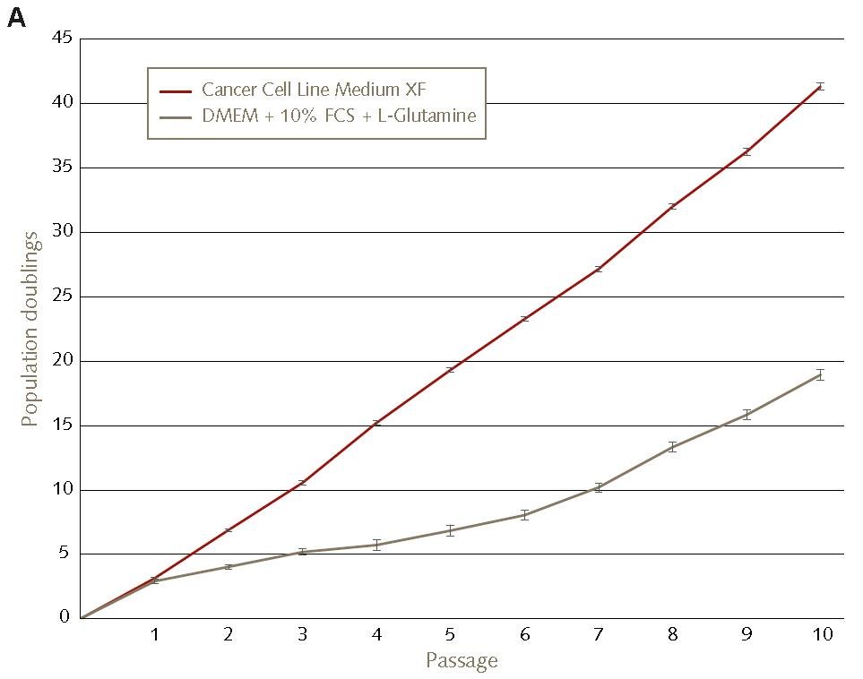
Figure 1. Accelerated growth kinetics of HT1080 fibrosarcoma cell lines in Promocell’s Cancer Cell Line Medium XF compared to conventional FBS containing culture conditions.HT1080 cells were plated with 5,000 cells/cm² in Cancer Cell Line Medium XF on fibronectin-coated vessels (red) or in DMEM + 2 mM L-Glutamine + 10% FCS (grey). Subsequently, the cells were cultured for 10 consecutive passages with a passage interval of 3 – 4 days.

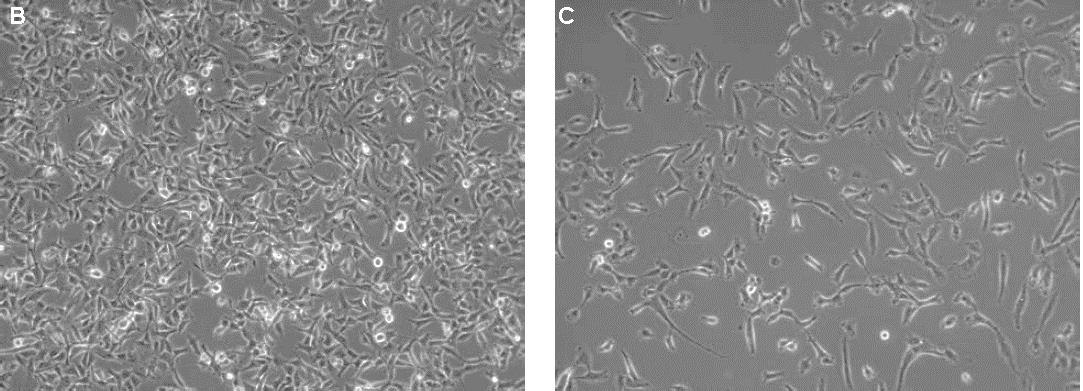
Figure 2. Morphology of HT1080 fibrosarcoma cells cultured in Cancer Cell Line Medium XF compared toconventional FBS containing culture conditions.HT1080 cells on day three after subculture (P7) are shown in the Cancer Cell Line Medium XF (B) and conventional FBS containing culture conditions (C) (100x magnification).

Figure 3. Morphology of MCF-7 breast carcinoma cells in the Cancer Cell Line Medium XF.MCF-7 cells were plated at 10,000 cells/cm2 in fibronectin-coated vessels and cultured for three passages in the Cancer Cell Line Medium XF. The cells exhibited efficient proliferation as well as a typical but slightly more compact compared to traditional culture media (not shown). Images were taken 1 day (A) and 5 days (B) after seeding at 100x magnification.
References
To continue reading please sign in or create an account.
Don't Have An Account?