Skip to Content
Merck
CN
All Photos(1)

Documents

Safety Information

MINCLARET

Sigma-Aldrich

CellVue® Claret Far Red Fluorescent Cell Linker Mini Kit for General Membrane Labeling

Distributed for Phanos Technologies

Sign Into View Organizational & Contract Pricing

Synonym(s):
Far red membrane labeling kit
UNSPSC Code:
12352207
NACRES:
NA.32

packaging

pkg of 1 kit

Quality Level

manufacturer/tradename

Distributed for Phanos Technologies

storage condition

protect from light

technique(s)

flow cytometry: suitable

fluorescence

λex 655 nm; λem 675 nm (CellVue claret dye)

application(s)

cell analysis
detection

detection method

fluorometric

shipped in

ambient

storage temp.

room temp

Related Categories

Application

CellVue Claret kit fluoresces in the far red and may be used for general cell membrane labeling including in vitro cell labeling, in vitro lymphocyte proliferation studies,† in vivo cytotoxicity assays,† and also as a second color for monitoring cell-cell membrane transfer.†
CellVue® Claret Far Red Fluorescent Cell Linker Mini Kit for General Membrane Labeling has been used:
  • to label monocyte-derived macrophages (MDMs) for antibody-dependent cellular phagocytosis assay
  • in labeling of Jurkat cells after the induction of apoptosi
  • in labeling of cells in mice for assaying the retention of tumor cells in lung

Biochem/physiol Actions

The CellVue Claret Fluorescent Cell Linker kit uses a proprietary membrane labeling technology to stably incorporate a far red fluorescent dye with long aliphatic tails (CellVue Claret) into lipid regions of the cell membrane.†

Packaging

This kit provides enough dye for up to 25 samples of 20 million cells each and enough diluent for up to 5 samples of 20 million cells each, when using a 2 mL final staining volume containing a final concentration of 2 x 10⁻⁶ M of PKH26 dye. Users must determine the optimal dye and cell concentrations from their cell type(s) and experimental purposes.

Linkage

For additional technical details on Fluorescent Cell Linker Dyes including a comprehensive bibliography, please visit PKH and CellVue®; Claret Fluorescent Cell Linker Dyes.

Legal Information

Distributed for Phanos Technologies, Inc.
Patent Information
CellVue is a registered trademark of Phanos Technologies

Pictograms

FlameExclamation mark

Signal Word

Danger

Hazard Statements

Hazard Classifications

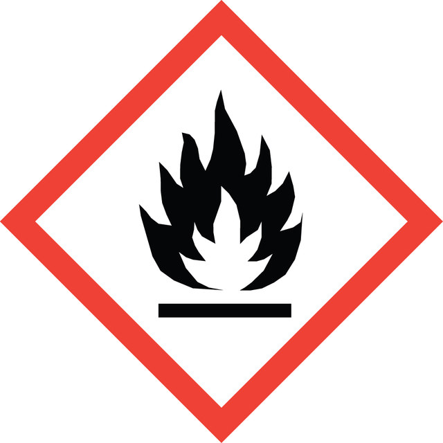
Eye Irrit. 2 - Flam. Liq. 2

WGK

WGK 1

Regulatory Information

危险化学品

Certificates of Analysis (COA)

Search for Certificates of Analysis (COA) by entering the products Lot/Batch Number. Lot and Batch Numbers can be found on a product’s label following the words ‘Lot’ or ‘Batch’.

Already Own This Product?

Find documentation for the products that you have recently purchased in the Document Library.

Visit the Document Library

Mitochondrial fission promotes the continued clearance of apoptotic cells by macrophages
Wang Y, et al.
Cell, 171(2), 331-345 (2017)
Comparative assessment of clinically utilized CD20-directed antibodies in chronic lymphocytic leukemia cells reveals divergent NK cell, monocyte, and macrophage properties
Rafiq S, et al.
Journal of immunology (Baltimore, Md. : 1950), 190(6), 2702-2711 (2013)
Roberts Rimsa et al.
Micromachines, 12(5) (2021-06-03)
Current in vitro models have significant limitations for new respiratory disease research and rapid drug repurposing. Lung on a chip (LOAC) technology offers a potential solution to these problems. However, these devices typically are fabricated from polydimethylsiloxane (PDMS), which has
The detergent-soluble cytoplasmic pool of survivin suppresses anoikis and its expression is associated with metastatic disease of human colon cancer
Hori M, et al.
Testing, 8(2), e55710-e55710 (2013)
Justin L Spinner et al.
Journal of leukocyte biology, 95(3), 389-398 (2013-11-15)
Yersinia pestis, the bacterial agent of plague, is transmitted by fleas. The bite of an infected flea deposits Y. pestis into the dermis and triggers recruitment of innate immune cells, including phagocytic PMNs. Y. pestis can subvert this PMN response

Articles

Lipophilic cell tracking dyes enable cancer biologists to track tumor and immune cell functions both in vitro and in vivo. Read the article to choose a right membrane dye kit for cell tracking and proliferation monitoring.

Optimal staining is a key component for studying tumorigenesis and progression. Learn useful tips and techniques for dye applications, including examples from recent studies.

Our team of scientists has experience in all areas of research including Life Science, Material Science, Chemical Synthesis, Chromatography, Analytical and many others.

Contact Technical Service