Skip to Content
Merck
CN
All Photos(1)

Key Documents

Safety Information

HPA005645

Sigma-Aldrich

Anti-GNB3 antibody produced in rabbit

Prestige Antibodies® Powered by Atlas Antibodies, affinity isolated antibody, buffered aqueous glycerol solution

Synonym(s):

Anti-Guanine nucleotide-binding protein G(I)/G(S)/G(T) subunit beta-3, Anti-Transducin beta chain 3

Sign Into View Organizational & Contract Pricing


About This Item

UNSPSC Code:
12352203
Human Protein Atlas Number:

biological source

rabbit

conjugate

unconjugated

antibody form

affinity isolated antibody

antibody product type

primary antibodies

clone

polyclonal

product line

Prestige Antibodies® Powered by Atlas Antibodies

form

buffered aqueous glycerol solution

species reactivity

human

technique(s)

immunohistochemistry (formalin-fixed, paraffin-embedded sections): suitable

immunogen sequence

ETGQQKTVFVGHTGDCMSLAVSPDFNLFISGACDASAKLWDVREGTCRQTFTGHESDINAICFFPNGEAICTGSDDASCRLFDLRADQELICFSHESIICGITSVAFSLSGRLLFAGYDDFNCNVWDSMKSERVGILSGHDNRVSC

UniProt accession no.

shipped in

wet ice

storage temp.

−20°C

Gene Information

human ... GNB3(2784)

Immunogen

Guanine nucleotide-binding protein G(I)/G(S)/G(T) subunit beta-3 recombinant protein epitope signature tag (PrEST)

Application

Applications in which this antibody has been used successfully, and the associated peer-reviewed papers, are given below.
Immunohistochemistry (1 paper)

Features and Benefits

Prestige Antibodies® are highly characterized and extensively validated antibodies with the added benefit of all available characterization data for each target being accessible via the Human Protein Atlas portal linked just below the product name at the top of this page. The uniqueness and low cross-reactivity of the Prestige Antibodies® to other proteins are due to a thorough selection of antigen regions, affinity purification, and stringent selection. Prestige antigen controls are available for every corresponding Prestige Antibody and can be found in the linkage section.

Every Prestige Antibody is tested in the following ways:
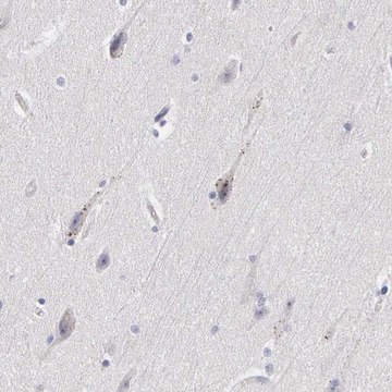
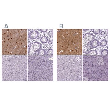
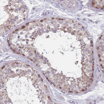
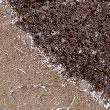
  • IHC tissue array of 44 normal human tissues and 20 of the most common cancer type tissues.
  • Protein array of 364 human recombinant protein fragments.

Physical form

Solution in phosphate-buffered saline, pH 7.2, containing 40% glycerol and 0.02% sodium azide

Legal Information

Prestige Antibodies is a registered trademark of Merck KGaA, Darmstadt, Germany

Disclaimer

Unless otherwise stated in our catalog or other company documentation accompanying the product(s), our products are intended for research use only and are not to be used for any other purpose, which includes but is not limited to, unauthorized commercial uses, in vitro diagnostic uses, ex vivo or in vivo therapeutic uses or any type of consumption or application to humans or animals.

Not finding the right product?  

Try our Product Selector Tool.

Storage Class Code

12 - Non Combustible Liquids

WGK

WGK 1

Flash Point(F)

Not applicable

Flash Point(C)

Not applicable

Personal Protective Equipment

dust mask type N95 (US), Eyeshields, Gloves

Regulatory Information

新产品

Choose from one of the most recent versions:

Certificates of Analysis (COA)

Lot/Batch Number

Don't see the Right Version?

If you require a particular version, you can look up a specific certificate by the Lot or Batch number.

Already Own This Product?

Find documentation for the products that you have recently purchased in the Document Library.

Visit the Document Library

Nicolás Cuenca et al.
Ophthalmology, 125(3), 407-422 (2017-10-19)
OCT has been a technological breakthrough in the diagnosis, treatment, and follow-up of many ocular diseases, especially retinal and neuro-ophthalmologic pathologic conditions. Until now, several controversies have arisen over the specific cell types that the bands observed in the OCT
Eric R Ritchey et al.
Experimental eye research, 102, 59-69 (2012-07-25)
Visual experience is known to guide ocular growth. We tested the hypothesis that vision-guided ocular growth is disrupted in a model system with diminished visual acuity. We examine whether ocular elongation is influenced by form-deprivation (FD) and lens-imposed defocus in
Shanti R Tummala et al.
The European journal of neuroscience, 43(11), 1509-1522 (2016-04-03)
Heterotrimeric G-proteins couple metabotropic receptors to downstream effectors. In retinal ON bipolar cells, Go couples the metabotropic receptor mGluR6 to the TRPM1 channel and closes it in the dark, thus hyperpolarizing the cell. Light, via GTPase-activating proteins, deactivates Go
Estrella Calvo et al.
International journal of molecular sciences, 21(5) (2020-02-29)
Combined administration of N-Methyl-D-Aspartate (NMDA) and kainic acid (KA) on the inner retina was studied as a model of excitotoxicity. The right eye of C57BL6J mice was injected with 1 µL of PBS containing NMDA 30 mM and KA 10
Sergei S Nikonov et al.
The Journal of neuroscience : the official journal of the Society for Neuroscience, 33(12), 5182-5194 (2013-03-22)
Mammalian cones respond to light by closing a cGMP-gated channel via a cascade that includes a heterotrimeric G-protein, cone transducin, comprising Gαt2, Gβ3 and Gγt2 subunits. The function of Gβγ in this cascade has not been examined. Here, we investigate

Our team of scientists has experience in all areas of research including Life Science, Material Science, Chemical Synthesis, Chromatography, Analytical and many others.

Contact Technical Service