DUO92014
Duolink® In Situ Detection Reagents Green
Sign In to View Organizational & Contract Pricing.
Select a Size
Change View
About This Item
NACRES:
NA.32
UNSPSC Code:
12352200
product line
Duolink®
Quality Segment
technique(s)
proximity ligation assay: suitable
fluorescence
λex 495 nm; λem 527 nm (green) (FITC (Cyanine 2), Zeiss Filter set 38)
suitability
suitable for fluorescence
shipped in
dry ice
storage temp.
−20°C
General description
Duolink® In Situ Detection Reagents Green contains all the necessary Duolink In situ reagents to perform the amplification and detection of bound PLA® probes. The detection probes contain a fluorophore (lex = 495 nm and lem = 527 nm), which may be visualized using the same filter as Cy®2 or FITC. Experiments conducted using Duolink In situ reagents can detect and visualize protein interactions, protein expression levels and post translational modifications at the single molecule level in fixed cells and tissue samples.
Application
Duolink®proximity ligation assay(PLA®) allows for endogenous detection of protein interactions, post translational modifications, and protein expression levels at the single molecule level in fixed cells and tissue samples.
Duolink® In Situ Detection Reagents has been used in the proximity ligation assay of:
- vasopressin and gonadotropin-releasing hormone (GnRH) from frozen rat brain sections
- human embryonic kidney 293 cells (HEK)
- pituitary tissues
Biochem/physiol Actions
Green fluorescence detection reagents are often used with FITC filter.
Features and Benefits
- No overexpression or genetic manipulation reNo overexpression or genetic manipulation required
- High specificity (fewer false positives)
- Single molecule sensitivity due to rolling circle amplification
- Relative quantification possible
- No special equipment needed
- Quicker and simpler than FRET
- Increased accuracy compared to co-IP
- Publication-ready results
Preparation Note
Store the components at –20 °C. The enzymes should be kept cold (–20 °C) at all times, use a freezing block when removing them from the freezer.
To perform a complete Duolink® PLA in situ experiment you will need two primary antibodies (PLA, IHC, ICC or IF validated) that recognize two target epitopes. Other necessary reagents include a pair of PLA probes from different species (one PLUS and one MINUS), detection reagents, wash buffers, and mounting medium. Note that the primary antibodies must come from the same species as the Duolink® PLA probes. Analysis is carried out using standard immunofluorescence assay equipment.
Other Notes
- 5x Ligation - Contains oligonucleotides that hybridize to the PLA probes and all components needed for ligation except the Ligase
- 1x Ligase (1 unit/μL)
- 1x Polymerase (10 units/μL)
- 5x Amplification Green - Contains all components needed for Rolling Circle Amplification (RCA) except the Polymerase. It also contains oligonucleotide probes labeled with a fluorophore that hybridize to the RCA product.
Not included in Detection kit:
Primary antibodies, PLA probes, wash buffers, mounting medium
Follow the Duolink® In Situ Fluorescence Protocol to use this product. A set of short instructionsis also available.
Visit our Duolink® PLA Resource Center for information on how to run a Duolink® experiment, applications, troubleshooting, and more.
Let us do the work for you, learn more about our Custom Service Program to accelerate your Duolink® projects
View full Duolink® product list
Visit our Duolink® PLA Resource Center for information on how to run a Duolink® experiment, applications, troubleshooting, and more.
Let us do the work for you, learn more about our Custom Service Program to accelerate your Duolink® projects
View full Duolink® product list
Legal Information
Cy is a registered trademark of Cytiva
Duolink is a registered trademark of Merck KGaA, Darmstadt, Germany
PLA is a registered trademark of Merck KGaA, Darmstadt, Germany
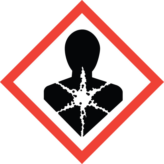
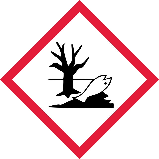
signalword
Danger
hcodes
Storage Class
10 - Combustible liquids
wgk
WGK 3
Hazard Classifications
Aquatic Chronic 2 - ED ENV 1 - Resp. Sens. 1
Choose from one of the most recent versions:
Already Own This Product?
Find documentation for the products that you have recently purchased in the Document Library.