
S7100
ApopTag Peroxidase In Situ Apoptosis Detection Kit
The ApopTag Peroxidase In Situ Apoptosis Detection Kit detects apoptotic cells in situ by labeling & detecting DNA strand breaks by the TUNEL method.
Synonym(s):
ApopTag Kit, In Situ Apoptosis Detection Kit
Select a Size
About This Item
Quality Level
manufacturer/tradename
ApopTag, Chemicon®
detection method
colorimetric
shipped in
dry ice
General description
Of all the aspects of apoptosis, the defining characteristic is a complete change in cellular morphology. As observed by electron microscopy, the cell undergoes shrinkage, chromatin margination, membrane blebbing, nuclear condensation and then segmentation, and division into apoptotic bodies which may be phagocytosed (11, 19, 24). The characteristic apoptotic bodies are short-lived and minute, and can resemble other cellular constituents when viewed by brightfield microscopy. DNA fragmentation in apoptotic cells is followed by cell death and removal from the tissue, usually within several hours (7). A rate of tissue regression as rapid as 25% per day can result from apparent apoptosis in only 2-3% of the cells at any one time (6). Thus, the quantitative measurement of an apoptotic index by morphology alone can be difficult.
DNA fragmentation is usually associated with ultrastructural changes in cellular morphology in apoptosis (26, 38). In a number of well-researched model systems, large fragments of 300 kb and 50 kb are first produced by endonucleolytic degradation of higher-order chromatin structural organization. These large DNA fragments are visible on pulsed-field electrophoresis gels (5, 43, 44). In most models, the activation of Ca2+- and Mg2+-dependent endonuclease activity further shortens the fragments by cleaving the DNA at linker sites between nucleosomes (3). The ultimate DNA fragments are multimers of about 180 bp nucleosomal units. These multimers appear as the familiar "DNA ladder" seen on standard agarose electrophoresis gels of DNA extracted from many kinds of apoptotic cells (e.g. 3, 7,13, 35, 44).
Another method for examining apoptosis via DNA fragmentation is by the TUNEL assay, (13) which is the basis of ApopTag technology. The DNA strand breaks are detected by enzymatically labeling the free 3′-OH termini with modified nucleotides. These new DNA ends that are generated upon DNA fragmentation are typically localized in morphologically identifiable nuclei and apoptotic bodies. In contrast, normal or proliferative nuclei, which have relatively insignificant numbers of DNA 3′-OH ends, usually do not stain with the kit. ApopTag Kits detect single-stranded (25) and double-stranded breaks associated with apoptosis. Drug-induced DNA damage is not identified by the TUNEL assay unless it is coupled to the apoptotic response (8). In addition, this technique can detect early-stage apoptosis in systems where chromatin condensation has begun and strand breaks are fewer, even before the nucleus undergoes major morphological changes (4, 8).
Apoptosis is distinct from accidental cell death (necrosis). Numerous morphological and biochemical differences that distinguish apoptotic from necrotic cell death are summarized in the following table (adapted with permission from reference 39).
Preparation Note
Precautions
1.The following kit components contain potassium cacodylate (dimethylarsinic acid) as a buffer: Equilibration Buffer (90416), Reaction Buffer (90417), and TdT Enzyme (90418). These components are harmful if swallowed; avoid contact with skin and eyes (wear gloves, glasses) and wash areas of contact immediately.
2. Antibody Conjugates (90420) and Blocking Solutions (#10 and #13) contain 0.08% sodium azide as a preservative.
3. TdT Enzyme (90418) contains glycerol and will not freeze at -20°C. For maximum shelf life, do not warm this reagent to room temp. before dispensing.
Other Notes
Reaction Buffer 2.0 mL -15°C to -25°C
TdT Enzyme 0.64 mL -15°C to -25°C
Stop/Wash Buffer 20 mL -15°C to -25°C
Anti-Digoxigenin-Peroxidase* 3.0 mL 2°C to 8°C
Plastic Coverslips 100 ea. Room Temp.
Note: Separate purchase of DAB (Peroxidase Substrate) is required. It is not supplied with this kit.
Number of samples per kit: Sufficient materials are provided to stain 40 tissue specimens of approximately 5 mm2 each when used according to instructions. Reaction Buffer will be fully consumed before other reagents when kits are used for slide-mounted specimens.
Legal Information

signalword
Danger
hcodes
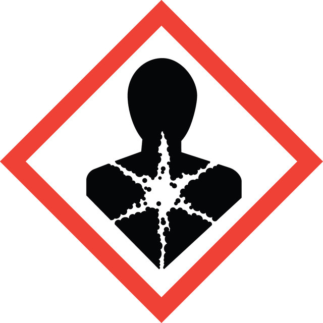
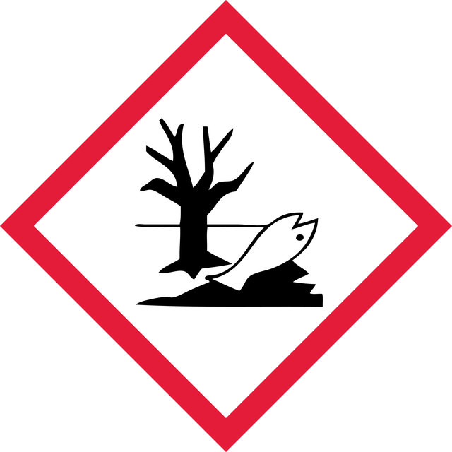
Hazard Classifications
Aquatic Chronic 2 - Carc. 1B - Skin Sens. 1 - STOT RE 2 Inhalation
target_organs
Respiratory Tract
Storage Class
6.1C - Combustible acute toxic Cat.3 / toxic compounds or compounds which causing chronic effects
wgk
WGK 3
Certificates of Analysis (COA)
Search for Certificates of Analysis (COA) by entering the products Lot/Batch Number. Lot and Batch Numbers can be found on a product’s label following the words ‘Lot’ or ‘Batch’.
Already Own This Product?
Find documentation for the products that you have recently purchased in the Document Library.
Articles
Cellular apoptosis assays to detect programmed cell death using Annexin V, Caspase and TUNEL DNA fragmentation assays.
细胞凋亡测定使用膜联蛋白V、Caspase和TUNEL DNA片段化检测法检测程序性细胞死亡。
Related Content
"Recognizing both the tremendous opportunities and the challenges facing cancer research, we are dedicated to developing and refining tools and technologies for the study of cancer. With our comprehensive portfolio, including the Upstate®, Chemicon®, and Calbiochem® brands of reagents and antibodies, researchers can count on dependable, high quality solutions for analyzing all the hallmarks of cancer."
Global Trade Item Number
| SKU | GTIN |
|---|---|
| S7100 | 04053252579356 |