Skip to Content
Merck
CN
All Photos(1)

Key Documents

OP43

Sigma-Aldrich

Anti-p53 (Ab-6) (Pantropic) Mouse mAb (DO-1)

liquid, clone DO-1, Calbiochem®

Synonym(s):

Anti-p53 Antibody, Mouse Anti-p53, p53 Detection Antibody

Sign Into View Organizational & Contract Pricing


About This Item

UNSPSC Code:
12352203
NACRES:
NA.43

biological source

mouse

Quality Level

antibody form

purified antibody

antibody product type

primary antibodies

clone

DO-1, monoclonal

form

liquid

contains

≤0.1% sodium azide as preservative

species reactivity

feline, human

should not react with

mouse, rat

manufacturer/tradename

Calbiochem®

storage condition

do not freeze

isotype

IgG2a

shipped in

wet ice

storage temp.

2-8°C

target post-translational modification

unmodified

Gene Information

human ... TP53(7157)

General description

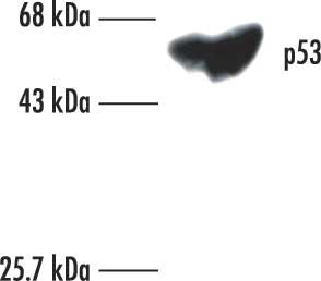
Purified mouse monoclonal antibody generated by immunizing BALB/c mice with the specified immunogen and fusing splenocytes with X63Ag8.653 mouse myeloma cells (see application references). Recognizes the ~53 kDa wild-type and mutant forms of p53.
Recognizes the ~53 kDa wild-type and mutant p53 protein in A431 cells and breast carcinoma tissue.
This Anti-p53 (Ab-6) (Pantropic) Mouse mAb (DO-1) is validated for use in Frozen sections, Immunoblotting, ICC, IP, Paraffin sections for the detection of p53 (Ab-6) (Pantropic).

Immunogen

Epitope: Within amino acids 21-25 of human p53
Human
wild type, recombinant, human p53

Application


Frozen sections (1 g/ml; see application references)
Immunoblotting (0.1-1 g/ml; see application references)
Immunocytochemistry (1-2.5 g/ml; see application references)
Immunoprecipitation (1 g/ml or use Cat. No. OP43A; see application references)
Paraffin sections (1 g/ml, pepsin, heat or pressure cooker pre-treatment required; see application references)

Packaging

Please refer to vial label for lot-specific concentration.

Warning

Toxicity: Standard Handling (A)

Physical form

In 0.05 M sodium phosphate buffer, 0.2% gelatin.

Analysis Note

Negative Control
SK-OV-3 cells or normal skin tissue
Positive Control
A431 cells or breast carcinoma tissue

Other Notes

El-Deiry, W.S., et al. 1994. Cancer Res.54, 1169.
Greenblatt, M.S., et al. 1994. Cancer Res.54, 4855.
Legros, Y., et al. 1994. Oncogene9, 2071.
Barak, Y., et al. 1993. EMBO J.12, 461.
Kastan, M.B., et al. 1992. Cell71, 587.
Kuerbitz, S.J. 1992. Proc. Natl. Acad. Sci. USA89, 7491.
Lane, D.P. 1992. Nature358, 15.
Vojtesek, B., et al. 1992. J. Immunol. Meth.151, 237.
Kastan, M.B., et al. 1991 Cancer Res.51, 6304.
Recognizes both mutant and wild-type p53 under denaturing and non-denaturing conditions. This antibody reacts weakly with rodent p53; we do not recommend it for rodent samples. For gel shift assay, use Cat. No. OP43L resuspended in 100 μl of buffer. Wild-type p53 has a short half life and is present in low amounts in cells. For immunoprecipitation increasing the amount of sample and labeling with 35S-Met for less than or equal to 1 h will aid in visualizing wild-type p53. Antibody should be titrated for optimal results in individual systems.

Legal Information

CALBIOCHEM is a registered trademark of Merck KGaA, Darmstadt, Germany

Not finding the right product?  

Try our Product Selector Tool.

Storage Class Code

10 - Combustible liquids

WGK

nwg

Flash Point(F)

Not applicable

Flash Point(C)

Not applicable


Certificates of Analysis (COA)

Search for Certificates of Analysis (COA) by entering the products Lot/Batch Number. Lot and Batch Numbers can be found on a product’s label following the words ‘Lot’ or ‘Batch’.

Already Own This Product?

Find documentation for the products that you have recently purchased in the Document Library.

Visit the Document Library

Yuji Masuda et al.
The Journal of biological chemistry, 294(11), 4177-4187 (2019-01-17)
Ubiquitin-specific protease 7 (USP7) regulates various cellular pathways through its deubiquitination activity. Despite the identification of a growing number of substrates of USP7, the molecular mechanism by which USP7 removes ubiquitin chains from polyubiquitinated substrates remains unexplored. The present study
Sadie Rice et al.
Viruses, 12(3) (2020-03-19)
Persistent infection by human papillomaviruses (HPVs), small, double-stranded DNA viruses that infect keratinocytes of the squamous epithelia, can lead to the development of cervical and other cancers. The viral oncoprotein E7 contributes to viral persistence in part by regulating host
Surendra Sharma et al.
Virology, 518, 8-13 (2018-02-11)
Modulation of expression of noncoding RNAs is an important aspect of the oncogenic activities of high-risk human papillomavirus (HPV) E6 and E7 proteins. While HPV E6/E7-mediated alterations of microRNAs (miRNAs) has been studied in detail there are fewer reports on
Dalton Dacus et al.
Journal of virology, 94(12) (2020-04-03)
Beta genus human papillomaviruses (β-HPVs) cause cutaneous squamous cell carcinomas (cSCCs) in a subset of immunocompromised patients. However, β-HPVs are not necessary for tumor maintenance in the general population. Instead, they may destabilize the genome in the early stages of
Rebecca A Dagg et al.
Cell reports, 19(12), 2544-2556 (2017-06-22)
Acquisition of replicative immortality is currently regarded as essential for malignant transformation. This is achieved by activating a telomere lengthening mechanism (TLM), either telomerase or alternative lengthening of telomeres, to counter normal telomere attrition. However, a substantial proportion of some

Our team of scientists has experience in all areas of research including Life Science, Material Science, Chemical Synthesis, Chromatography, Analytical and many others.

Contact Technical Service