Cell Imaging with Fluorescent Lentiviral Biosensors
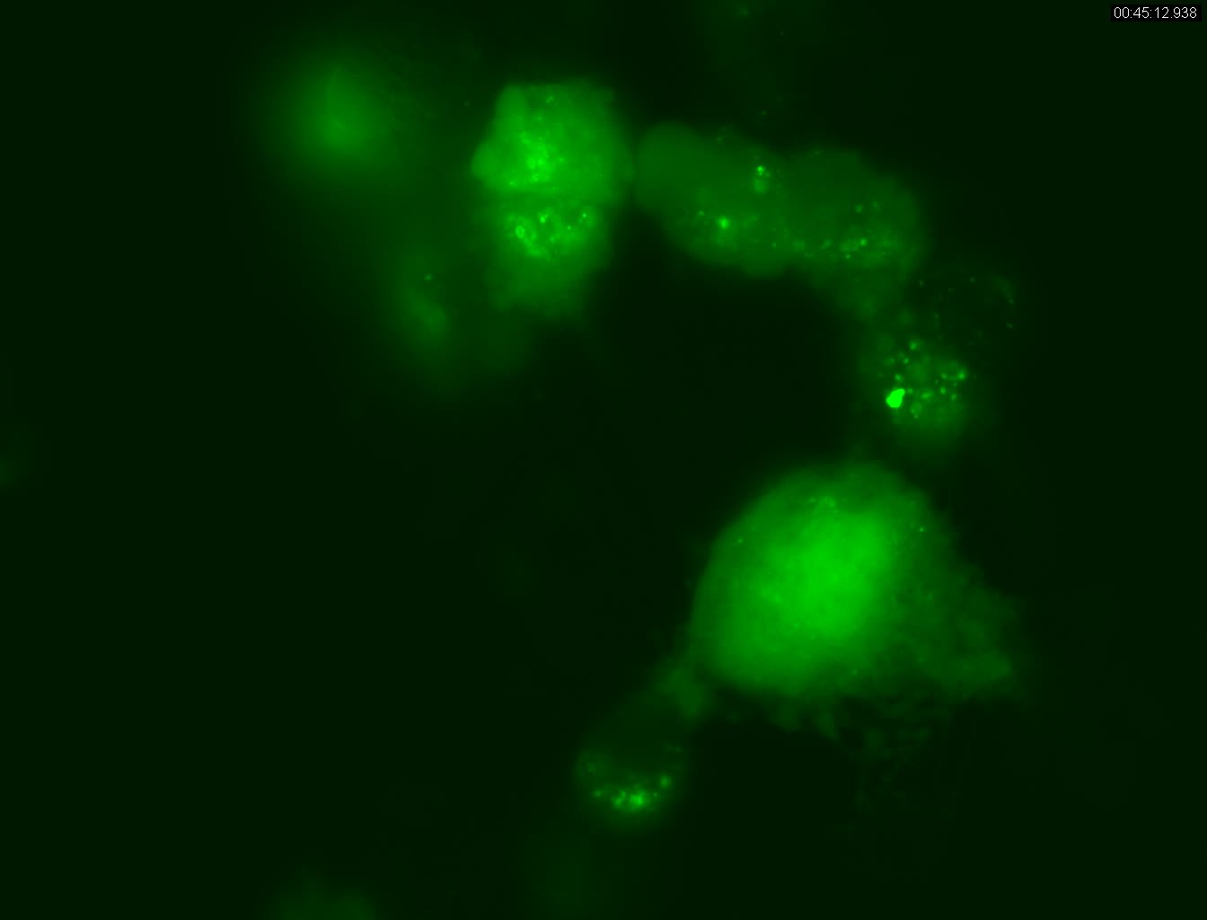
LentiBrite™ Live Cell Video Gallery
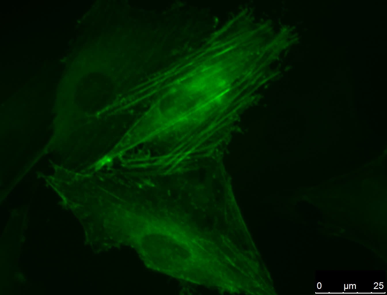
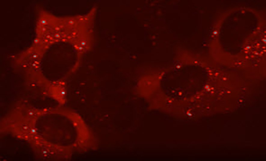
Biosensors can be used to detect a particular protein as well as the subcellular location of that protein within live cells. Fluorescent tags such as GFP and RFP are an effective way to visualize the protein of interest within a cell by either fluorescent microscopy or time-lapse video capture. Visualizing live cells without disruption can reveal changing cellular conditions in real time. Lentiviral vector systems are a popular tool for introducing genes and gene products into cells. Advantages over non-viral methods (such as chemical-based transfection) include higher-efficiency transfection of dividing and non-dividing cells, stable expression of the transgene, and low immunogenicity.
LentiBrite™ Fluorescent Lentiviral Biosensors are a new suite of pre-packaged lentiviral particles encoding important and foundational proteins of autophagy, apoptosis, and cell structure that enables visualization under different cell/disease states in live cell and in vitro analysis.
Features and Benefits
- Pre-packaged, ready-to-use, fluorescently-tagged with monomeric GFP & RFP
- Minimum titer (≥3 x 10⁸ IFU/mL) per vial
- Higher efficiency transfection as compared to traditional chemical-based and other non-viral-based transfection methods
- Ability to transfect dividing, non-dividing, and difficult-to-transfect cell types, such as primary cells or stem cells
- Non-disruptive towards cellular function
- Validated for fluorescent microscopy and live cell analysis
Materials
To continue reading please sign in or create an account.
Don't Have An Account?

