Abberior® Dyes for Super-Resolution Microscopy Applications
Microscopic methods in life sciences are of tremendous importance for visualization cellular and tissue structures. The development of new microscopy concepts has overcome the resolution barrier given by the diffraction limit, enabling a resolution limit down to about 10 nm. The visualization of cellular structures and molecular interactions can reveal new understanding in biological processes. Due to tremendous efforts in the development of super-resolved fluorescence microscopy, The Nobel Prize in Chemistry for 2014 was awarded to Eric Betzig, Stefan W. Hell, and to William E. Moerner.
These super-resolution microscopy principles are based on several technological approaches. Conventional light microscopy enables a resolution limit of about 250 nm in the x- and y- direction and 450 – 700 nm in the z –direction. Super-resolution techniques have overcome the resolution-limit (point-spread function) at least by a factor of 2. The resolution of super-resolution microscopy depends on the number of points that can be resolved on the structure of interest. Crucial for a successful super-resolution imaging is the choice of fluorescent probe. Brightness and high-contrast ratio between the states are important. In most super-resolution methods, the states of the probe must be controllable, reversible or irreversible, and switchable between a light or a dark state. Depending on the super-resolution method, further photo-physical criteria the probe must be fulfilled. Established techniques include the following:
- STED (Stimulated emission depletion)
- GSDIM (Ground State Depletion)
- PALM (Photoactivated localization microscopy)
- STORM (Stochastic optical reconstruction microscopy)
- RESOLFT (reversible saturable optical (flurorescence) transitions)

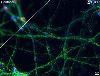
Figure 1.Three-color STED image of primary hippocampal neurons imaged with the Abberior® Instruments Expert Line STED microscope. Note: The characteristic ~190 nm beta II spectrin periodicity along distal axons (green), which is only visible in the STED image. Labeled structures and dyes: beta II Spectrin (green, Abberior® STAR580), Bassoon (red, Abberior® STAR635P), Actin cytoskeleton (blue, Phalloidin, Oregon Green 488). Sample was prepared by Elisa D’Este at MPIBPC, Göttingen.
We offer the superior series of Abberior® dyes that are especially designed and tested for super-resolution microscopy such as STED, RESOLFT, PALM, STORM, GSDIM and others. Abberior® STAR, Abberior® CAGE, Abberior® FLIP, Abberior® RSFP – the specific requirements of the super-resolution techniques are served with dedicated dye series. These Dyes are developed and produced by Abberior® GmbH. Stefan Hell is it’s Co-founder.
Benefits
- Optimized for brightness and low background
- Optimized switching behavior being the key for super-resolution
- All markers are tested for different super-resolution methods
- Abberior® STAR for STED, confocal and epifluorescence imaging
- Abberior® CAGE & FLIP for PALM, STORM and GSDIM
- Abberior® dyes are recommended by microscope vendors
- Proprietary, IP protected products
- Detailed characteristics of the dyes provided, e.g. optimal STED wavelength
Super-resolution microscopy is dependent on fluorescent labels. Manufactured by Abberior®, the STAR, CAGE and FLIP dyes as well as RSFPs are exceptionally bright and photostable and provide optimized photoswitching for RESOLFT and PALM/STORM imaging. They are the only commercially available dyes that are tailored specifically to the needs of super-resolution microscopy.
Abberior® dyes are also well suited for confocal microscopy, epifluorescence imaging and single molecule applications. Fluorescence applications that depend on a good signal-to-noise ratios and low background cam benefit from Abberior® dyes.

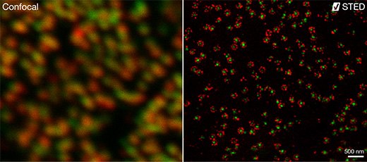
Confocal and STED images. Two subunits of the nuclear pore complex were immunolabeled using antibodies against gp210 and antibodies with multiple specificities (PAN4/5) and secondary antibodies coupled to Abberior® STAR580 and Abberior® STAR635P. Note that gp210 is localized in an eight-fold symmetric structure at the rim of the nuclear pore complex. Imaged with the Abberior® Instruments STEDYCON (compact line).
For North America, we also provide a series of antibody labeled Abberior® conjugates.
References
To continue reading please sign in or create an account.
Don't Have An Account?