Skip to Content
Merck
CN
All Photos(6)

Documents

Safety Information

HPA022962

Sigma-Aldrich

Anti-DCAF7 antibody produced in rabbit

enhanced validation

Prestige Antibodies® Powered by Atlas Antibodies, affinity isolated antibody, buffered aqueous glycerol solution, Ab2

Sign Into View Organizational & Contract Pricing

Synonym(s):
Anti-WD repeat-containing protein 68, Anti-WD repeat-containing protein An11 homolog, Anti-WDR68
UNSPSC Code:
12352203
Human Protein Atlas Number:
NACRES:
NA.41

biological source

rabbit

Quality Level

conjugate

unconjugated

antibody form

affinity isolated antibody

antibody product type

primary antibodies

clone

polyclonal

product line

Prestige Antibodies® Powered by Atlas Antibodies

form

buffered aqueous glycerol solution

species reactivity

human

enhanced validation

recombinant expression
independent
Learn more about Antibody Enhanced Validation

technique(s)

immunoblotting: 0.04-0.4 μg/mL
immunohistochemistry: 1:20-1:50

immunogen sequence

YPDLLATSGDYLRVWRVGETETRLECLLNNNKNSDFCAPLTSFDWNEVDPYLLGTSSIDTTCTIWGLETGQVLGRVNLVSGHVKTQ

shipped in

wet ice

storage temp.

−20°C

target post-translational modification

unmodified

Gene Information

human ... WDR68(10238)

General description

DCAF7 (DDB1 and CUL4 associated factor 7) is a WD40 (β-transducin) repeat containing protein. It is crucial for the survival of mammalian cells and facilitates protein-protein interaction. It interacts with various proteins, such as dual specificity tyrosine phosphorylation-regulated protein kinase (DYRK1A), MAPK/ERK kinase kinase 1 (MEKK1), and Cullin4-damage-specific DNA-binding protein 1 (CUL4-DDB1). The gene is mapped to human chromosome 17q23.

Immunogen

WD repeat-containing protein 68 recombinant protein epitope signature tag (PrEST)

Application

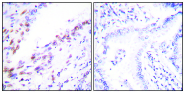
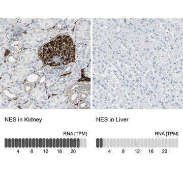
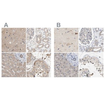
Anti-DCAF7 antibody produced in rabbit, a Prestige Antibody, is developed and validated by the Human Protein Atlas (HPA) project . Each antibody is tested by immunohistochemistry against hundreds of normal and disease tissues. These images can be viewed on the Human Protein Atlas (HPA) site by clicking on the Image Gallery link. The antibodies are also tested using immunofluorescence and western blotting. To view these protocols and other useful information about Prestige Antibodies and the HPA, visit sigma.com/prestige.
Applications in which this antibody has been used successfully, and the associated peer-reviewed papers, are given below.
Immunofluorescence (1 paper)

Features and Benefits

Prestige Antibodies® are highly characterized and extensively validated antibodies with the added benefit of all available characterization data for each target being accessible via the Human Protein Atlas portal linked just below the product name at the top of this page. The uniqueness and low cross-reactivity of the Prestige Antibodies® to other proteins are due to a thorough selection of antigen regions, affinity purification, and stringent selection. Prestige antigen controls are available for every corresponding Prestige Antibody and can be found in the linkage section.

Every Prestige Antibody is tested in the following ways:
  • IHC tissue array of 44 normal human tissues and 20 of the most common cancer type tissues.
  • Protein array of 364 human recombinant protein fragments.

Linkage

Corresponding Antigen APREST75709

Physical form

Solution in phosphate-buffered saline, pH 7.2, containing 40% glycerol and 0.02% sodium azide

Legal Information

Prestige Antibodies is a registered trademark of Merck KGaA, Darmstadt, Germany

WGK

WGK 1

Flash Point(F)

Not applicable

Flash Point(C)

Not applicable

Regulatory Information

新产品

Certificates of Analysis (COA)

Search for Certificates of Analysis (COA) by entering the products Lot/Batch Number. Lot and Batch Numbers can be found on a product’s label following the words ‘Lot’ or ‘Batch’.

Already Own This Product?

Find documentation for the products that you have recently purchased in the Document Library.

Visit the Document Library

Fabrice Sircoulomb et al.
EMBO molecular medicine, 3(3), 153-166 (2011-02-18)
Luminal B breast cancers represent a fraction of oestrogen receptor (ER)-positive tumours associated with poor recurrence-free and disease-specific survival in all adjuvant systemic treatment categories including hormone therapy alone. Identification of specific signalling pathways driving luminal B biology is paramount
Elizabeth J Leslie et al.
Human molecular genetics, 25(13), 2862-2872 (2016-04-02)
Orofacial clefts (OFCs), which include non-syndromic cleft lip with or without cleft palate (CL/P), are among the most common birth defects in humans, affecting approximately 1 in 700 newborns. CL/P is phenotypically heterogeneous and has a complex etiology caused by
Yoshihiko Miyata et al.
The Journal of biological chemistry, 289(48), 33320-33332 (2014-10-25)
Trp-Asp (WD) repeat protein 68 (WDR68) is an evolutionarily conserved WD40 repeat protein that binds to several proteins, including dual specificity tyrosine phosphorylation-regulated protein kinase (DYRK1A), MAPK/ERK kinase kinase 1 (MEKK1), and Cullin4-damage-specific DNA-binding protein 1 (CUL4-DDB1). WDR68 affects multiple

Our team of scientists has experience in all areas of research including Life Science, Material Science, Chemical Synthesis, Chromatography, Analytical and many others.

Contact Technical Service