Skip to Content
Merck
CN
All Photos(2)

Documents

Safety Information

HPA018468

Sigma-Aldrich

Anti-GDF3 antibody produced in rabbit

enhanced validation

Prestige Antibodies® Powered by Atlas Antibodies, affinity isolated antibody, buffered aqueous glycerol solution

Sign Into View Organizational & Contract Pricing

Synonym(s):
Anti-GDF-3, Anti-Growth/differentiation factor 3
UNSPSC Code:
12352203
Human Protein Atlas Number:
NACRES:
NA.41

biological source

rabbit

Quality Level

conjugate

unconjugated

antibody form

affinity isolated antibody

antibody product type

primary antibodies

clone

polyclonal

product line

Prestige Antibodies® Powered by Atlas Antibodies

form

buffered aqueous glycerol solution

species reactivity

human

enhanced validation

recombinant expression
Learn more about Antibody Enhanced Validation

technique(s)

immunoblotting: 0.04-0.4 μg/mL
immunohistochemistry: 1:20-1:50

immunogen sequence

HKWIIAPKGFMANYCHGECPFSLTISLNSSNYAFMQALMHAVDPEIPQAVCIPTKLSPISMLYQDNNDNVILRHYEDMVV

UniProt accession no.

shipped in

wet ice

storage temp.

−20°C

target post-translational modification

unmodified

Gene Information

human ... GDF3(9573)

General description

The growth differentiation factor-3 (GDF3) gene is mapped to human chromosome 12p13.1. It belongs to the bone morphogenic protein/GDF class of the transforming growth factor-β superfamily. GDF3 is expressed in embryonic stem cells and the early embryo. It is also detected in primary hippocampal neurons, and brain tissues. The protein is localized in the cytoplasm.

Immunogen

Growth/differentiation factor 3 Precursor recombinant protein epitope signature tag (PrEST)

Application

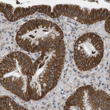
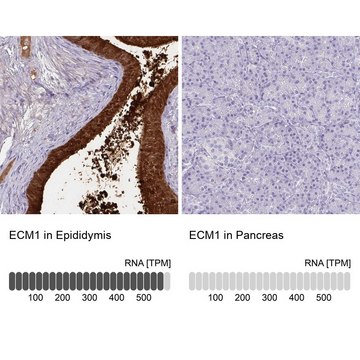
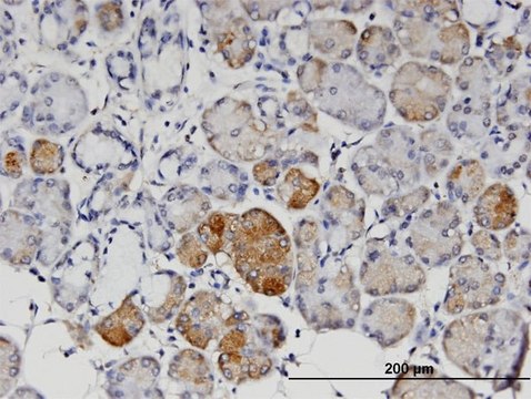
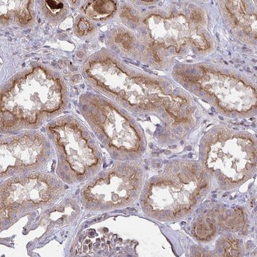
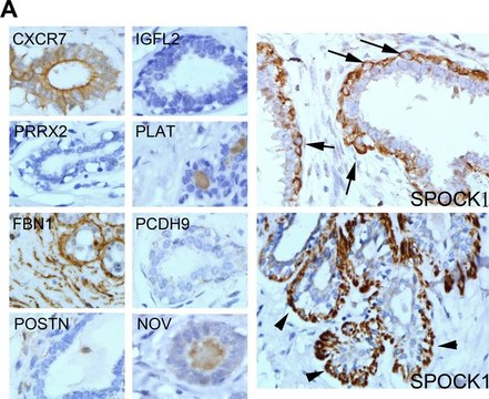
All Prestige Antibodies Powered by Atlas Antibodies are developed and validated by the Human Protein Atlas (HPA) project and as a result, are supported by the most extensive characterization in the industry.

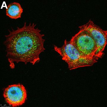
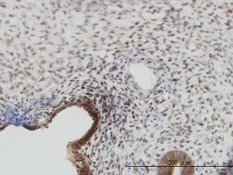
The Human Protein Atlas project can be subdivided into three efforts: Human Tissue Atlas, Cancer Atlas, and Human Cell Atlas. The antibodies that have been generated in support of the Tissue and Cancer Atlas projects have been tested by immunohistochemistry against hundreds of normal and disease tissues and through the recent efforts of the Human Cell Atlas project, many have been characterized by immunofluorescence to map the human proteome not only at the tissue level but now at the subcellular level. These images and the collection of this vast data set can be viewed on the Human Protein Atlas (HPA) site by clicking on the Image Gallery link. We also provide Prestige Antibodies® protocols and other useful information.

Biochem/physiol Actions

In embryonic stem (ES) cells growth/differentiation factor-3 (GDF3) is involved in maintaining the undifferentiated state and the ability to differentiate into the full spectrum of cell types. Bone morphogenetic protein 6 (BMP-6) and BMP-7 induces expression of GDF3. Presence of GDF3 increases luteinizing hormone receptor mRNA expression in human granulosa cells. GDF3 is highly expressed in adipose tissue. It regulates adipose-tissue homeostasis and diet-induced obesity under a high lipid load. GDF3 participates in ocular and skeletal development. Mutation in GDF3 causes ocular and skeletal anomalies.

Features and Benefits

Prestige Antibodies® are highly characterized and extensively validated antibodies with the added benefit of all available characterization data for each target being accessible via the Human Protein Atlas portal linked just below the product name at the top of this page. The uniqueness and low cross-reactivity of the Prestige Antibodies® to other proteins are due to a thorough selection of antigen regions, affinity purification, and stringent selection. Prestige antigen controls are available for every corresponding Prestige Antibody and can be found in the linkage section.

Every Prestige Antibody is tested in the following ways:
  • IHC tissue array of 44 normal human tissues and 20 of the most common cancer type tissues.
  • Protein array of 364 human recombinant protein fragments.

Linkage

Corresponding Antigen APREST74088

Physical form

Solution in phosphate-buffered saline, pH 7.2, containing 40% glycerol and 0.02% sodium azide

Legal Information

Prestige Antibodies is a registered trademark of Merck KGaA, Darmstadt, Germany

WGK

WGK 1

Flash Point(F)

Not applicable

Flash Point(C)

Not applicable

Regulatory Information

新产品

Certificates of Analysis (COA)

Search for Certificates of Analysis (COA) by entering the products Lot/Batch Number. Lot and Batch Numbers can be found on a product’s label following the words ‘Lot’ or ‘Batch’.

Already Own This Product?

Find documentation for the products that you have recently purchased in the Document Library.

Visit the Document Library

Ming Ye et al.
Human molecular genetics, 19(2), 287-298 (2009-10-30)
Ocular mal-development results in heterogeneous and frequently visually disabling phenotypes that include coloboma and microphthalmia. Due to the contribution of bone morphogenetic proteins to such processes, the function of the paralogue Growth Differentiation Factor 3 was investigated. Multiple mis-sense variants
Olov Andersson et al.
Proceedings of the National Academy of Sciences of the United States of America, 105(20), 7252-7256 (2008-05-16)
Growth/differentiation factor 3 (GDF3) is highly expressed in adipose tissue, and previous overexpression experiments in mice have suggested that it may act as an adipogenic factor under conditions of high lipid load. GDF3 has been shown to signal via the
Jia Shi et al.
Fertility and sterility, 97(4), 979-983 (2012-02-07)
To examine the relevance of growth differentiation factor 3 (GDF-3) and bone morphogenetic protein (BMP) cytokines in human ovary. Molecular studies. Research laboratory. Eight women undergoing salpingo-oophorectomy and 30 women undergoing ovarian stimulation for in vitro fertilization. Localizing GDF-3 protein in
Qiang Li et al.
Molecular and cellular biochemistry, 359(1-2), 115-123 (2011-08-02)
Members of the transforming growth factor-β (TGF-β) superfamily have significant roles in the regulation of a wide variety of physiological processes. In our present work, phylogenetic tree analysis showed that human GDF3 (Growth and differentiation factor 3) and human GDF1
Sung-Won Park et al.
FEBS letters, 586(19), 3529-3535 (2012-09-12)
We investigated transactivation by NANOG in regulating growth and differentiation factor 3 (GDF3) expression in NCCIT cells. GDF3 expression was affected by shRNA-mediated downregulation and by exogenous overexpression of NANOG specifically, as well as by retinoic acid-mediated differentiation. GDF3 transcription

Our team of scientists has experience in all areas of research including Life Science, Material Science, Chemical Synthesis, Chromatography, Analytical and many others.

Contact Technical Service