Skip to Content
Merck
CN
All Photos(7)

Key Documents

Safety Information

HPA006723

Sigma-Aldrich

Anti-LIG3 antibody produced in rabbit

Prestige Antibodies® Powered by Atlas Antibodies, affinity isolated antibody, buffered aqueous glycerol solution

Synonym(s):

Anti-DNA ligase 3, Anti-DNA ligase III, Anti-Polydeoxyribonucleotide synthase [ATP] 3

Sign Into View Organizational & Contract Pricing


About This Item

UNSPSC Code:
12352203
Human Protein Atlas Number:

biological source

rabbit

conjugate

unconjugated

antibody form

affinity isolated antibody

antibody product type

primary antibodies

clone

polyclonal

product line

Prestige Antibodies® Powered by Atlas Antibodies

form

buffered aqueous glycerol solution

species reactivity

human

technique(s)

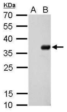
immunoblotting: 0.04-0.4 μg/mL
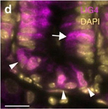
immunofluorescence: 0.25-2 μg/mL
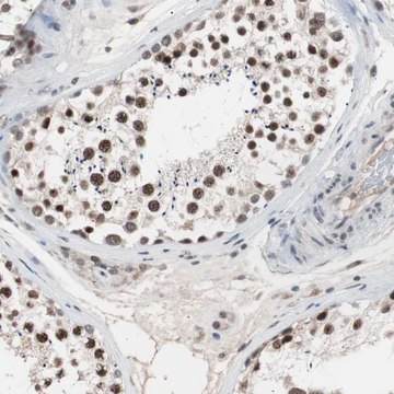
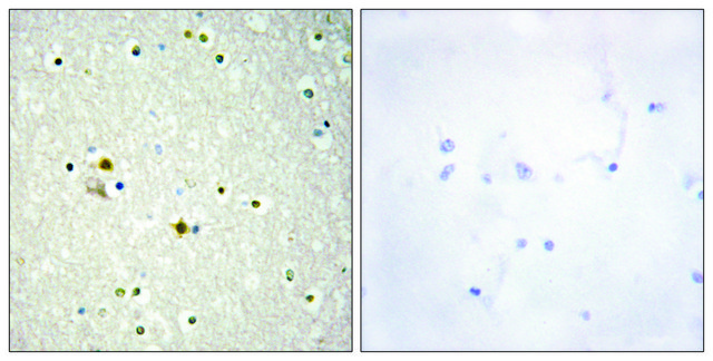
immunohistochemistry: 1:50-1:200

immunogen sequence

CDPRHKDCLLREFRKLCAMVADNPSYNTKTQIIQDFLRKGSAGDGFHGDVYLTVKLLLPGVIKTVYNLNDKQIVKLFSRIFNCNPDDMARDLEQGDVSETIRVFFEQSKSFPPAAKSLLTIQEVDEFLLRLSKLTKE

UniProt accession no.

shipped in

wet ice

storage temp.

−20°C

target post-translational modification

unmodified

Gene Information

human ... LIG3(3980)

Immunogen

DNA ligase 3 recombinant protein epitope signature tag (PrEST)

Application

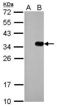
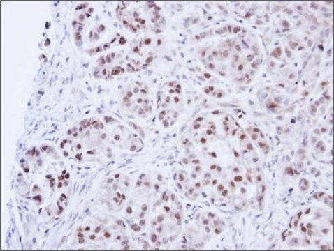
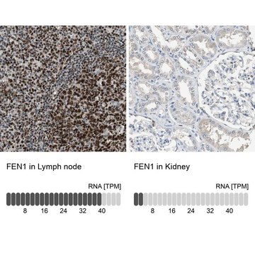
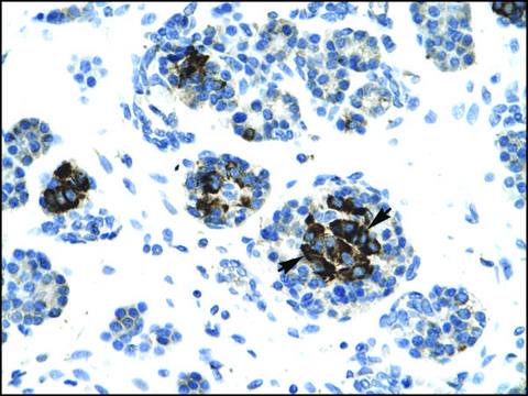
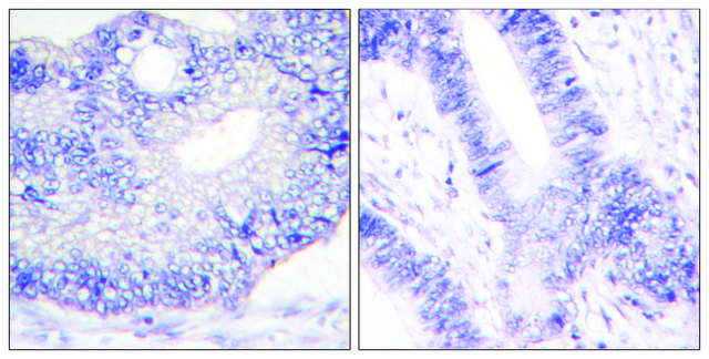
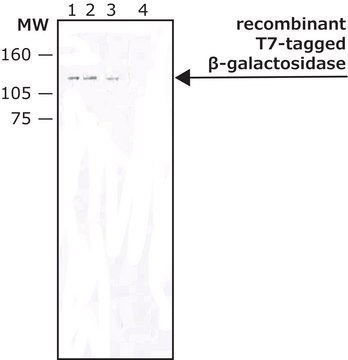
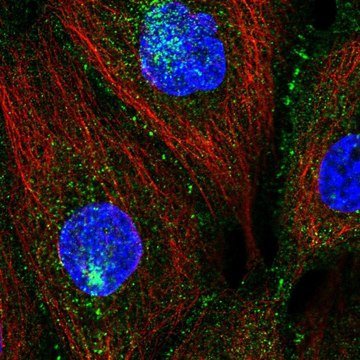
Anti-LIG3 antibody produced in rabbit, a Prestige Antibody, is developed and validated by the Human Protein Atlas (HPA) project . Each antibody is tested by immunohistochemistry against hundreds of normal and disease tissues. These images can be viewed on the Human Protein Atlas (HPA) site by clicking on the Image Gallery link. The antibodies are also tested using immunofluorescence and western blotting. To view these protocols and other useful information about Prestige Antibodies and the HPA, visit sigma.com/prestige.

Biochem/physiol Actions

LIG3 (Ligase 3) is a DNA repair protein comprises of a PARP-like zinc finger (ZnF) domain and OB-fold (OBD) domain. It plays a key role in nuclear and mitochondrial DNA replication and repair. The ZnF-DNA binding domain first forms a crescent-shaped surface to identify the DNA end part. Later, it catalyzes the repair with the help of nucleotidyl transferase (NTase) and OB-fold (OBD) domain. It identifies nick site and increases the extent of DNA nick joining followed by intermolecular DNA ligation. In human mitochondrial replication system, LIG3 joins the Okazaki fragment at the DNA break site.

Features and Benefits

Prestige Antibodies® are highly characterized and extensively validated antibodies with the added benefit of all available characterization data for each target being accessible via the Human Protein Atlas portal linked just below the product name at the top of this page. The uniqueness and low cross-reactivity of the Prestige Antibodies® to other proteins are due to a thorough selection of antigen regions, affinity purification, and stringent selection. Prestige antigen controls are available for every corresponding Prestige Antibody and can be found in the linkage section.

Every Prestige Antibody is tested in the following ways:
  • IHC tissue array of 44 normal human tissues and 20 of the most common cancer type tissues.
  • Protein array of 364 human recombinant protein fragments.

Linkage

Corresponding Antigen APREST71048

Physical form

Solution in phosphate-buffered saline, pH 7.2, containing 40% glycerol and 0.02% sodium azide

Legal Information

Prestige Antibodies is a registered trademark of Merck KGaA, Darmstadt, Germany

Disclaimer

Unless otherwise stated in our catalog or other company documentation accompanying the product(s), our products are intended for research use only and are not to be used for any other purpose, which includes but is not limited to, unauthorized commercial uses, in vitro diagnostic uses, ex vivo or in vivo therapeutic uses or any type of consumption or application to humans or animals.

Not finding the right product?  

Try our Product Selector Tool.

Storage Class Code

10 - Combustible liquids

WGK

WGK 1

Flash Point(F)

Not applicable

Flash Point(C)

Not applicable

Personal Protective Equipment

dust mask type N95 (US), Eyeshields, Gloves

Regulatory Information

常规特殊物品

Choose from one of the most recent versions:

Certificates of Analysis (COA)

Lot/Batch Number

Don't see the Right Version?

If you require a particular version, you can look up a specific certificate by the Lot or Batch number.

Already Own This Product?

Find documentation for the products that you have recently purchased in the Document Library.

Visit the Document Library

Heini Ruhanen et al.
Biochimica et biophysica acta, 1813(12), 2000-2007 (2011-09-01)
Recent evidence suggests that coupled leading and lagging strand DNA synthesis operates in mammalian mitochondrial DNA (mtDNA) replication, but the factors involved in lagging strand synthesis are largely uncharacterised. We investigated the effect of knockdown of the candidate proteins in
Elizabeth Cotner-Gohara et al.
Biochemistry, 49(29), 6165-6176 (2010-06-04)
Human DNA ligase III has essential functions in nuclear and mitochondrial DNA replication and repair and contains a PARP-like zinc finger (ZnF) that increases the extent of DNA nick joining and intermolecular DNA ligation, yet the bases for ligase III
Jinshui Fan et al.
Blood, 116(24), 5298-5305 (2010-09-03)
The internal tandem duplication (ITD) mutations of the FMS-like tyrosine kinase-3 (FLT3) receptor found in acute myeloid leukemia patients are associated with poor prognosis. Although DNA double-strand breaks (DSBs) are mainly repaired by the DNA-PK-dependent nonhomologous end-joining (NHEJ) pathway in
Elena M Cortizas et al.
Journal of immunology (Baltimore, Md. : 1950), 191(11), 5751-5763 (2013-10-23)
Classical nonhomologous end-joining (C-NHEJ) and alternative end-joining (A-EJ) are the main DNA double-strand break (DSB) repair pathways when a sister chromatid is not available. However, it is not clear how one pathway is chosen over the other to process a
Roland Steinacher et al.
The EMBO journal, 38(1) (2018-12-14)
During active DNA demethylation, 5-methylcytosine (5mC) is oxidized by TET proteins to 5-formyl-/5-carboxylcytosine (5fC/5caC) for replacement by unmethylated C by TDG-initiated DNA base excision repair (BER). Base excision generates fragile abasic sites (AP-sites) in DNA and has to be coordinated

Our team of scientists has experience in all areas of research including Life Science, Material Science, Chemical Synthesis, Chromatography, Analytical and many others.

Contact Technical Service