
OP64
Anti-p21WAF1 (Ab-1) Mouse mAb (EA10)
liquid, clone EA10, Calbiochem®
Synonym(s):
Anti-CIP1, Anti-SD11, Anti-p21, Anti-WAF
Select a Size
About This Item
biological source
mouse
Quality Level
antibody form
purified antibody
antibody product type
primary antibodies
clone
EA10, monoclonal
form
liquid
contains
≤0.1% sodium azide as preservative
species reactivity
human
should not react with
mouse, rat
manufacturer/tradename
Calbiochem®
storage condition
do not freeze
dilution
(Flow Cytometry (2 µg/mL)
Frozen Sections (5 µg/mL)
Immunoblotting (1-3 µg/mL)
Immunofluorescence (1-5 µg/mL)
Immunoprecipitation (2 µg/sample)
Paraffin Sections (5 µg/mL or use OP64F; heat pre-treatment required; and application references))
isotype
IgG1
shipped in
wet ice
storage temp.
2-8°C
target post-translational modification
unmodified
Gene Information
human ... CDKN1A(1026)
General description
Application
Flow Cytometry (2 g/ml or use Cat. No. OP64F; see application references)
Frozen Sections (5 g/ml or use Cat. No. OP64F)
Immunoblotting (1-3 g/ml)
Immunofluorescence (1-5 g/ml or use Cat. No. OP64F)
Immunoprecipitation (2 g/sample)
Paraffin Sections (5 g/ml or use OP64F; heat pre-treatment required; see comments and application references)
Packaging
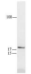
Analysis Note
Any cell line expressing wild-type p53 (e.g. Hs27 or U2OS treated with DNA-damaging agents) or skin or colon tissue
Other Notes
Chen, Y.Q., et al. 1995. Int. J. Oncology7, 889.
Deng, C., et al. 1995. Cell82, 675.
El-Deiry, W.S., et al. 1995. Cancer Res.55, 2910.
Waldman, T., et al. 1995. Cancer Res.55, 5187.
Elbendary, A., et al.1994. Cell Growth Diff.5, 1301.
El-Deiry, W.S., et al. 1994 Cancer Res.54, 1169.
Li, R., et al. 1994. Nature371, 534.
Michieli, P., et al. 1994. Cancer Res.54, 3391.
Noda, A., et al.1994. Exp. Cell Res.211, 90.
El-Deiry, W.S., et al.1993. Cell75, 817.
Gu, Y., et al. 1993. Nature366, 707.
Harper, J.W., et al.1993. Cell75, 805.
Xiong, Y., et al.1993. Genes Devel.7, 1572.
Xiong, Y., et al.1993. Nature366, 701.
Xiong, Y., et al.1992. Cell71, 505.
Legal Information
Disclaimer
Still not finding the right product?
Try our Product Selector Tool to narrow your options
Storage Class
11 - Combustible Solids
wgk
WGK 1
flash_point_f
Not applicable
flash_point_c
Not applicable
Certificates of Analysis (COA)
Search for Certificates of Analysis (COA) by entering the products Lot/Batch Number. Lot and Batch Numbers can be found on a product’s label following the words ‘Lot’ or ‘Batch’.
Already Own This Product?
Find documentation for the products that you have recently purchased in the Document Library.
Global Trade Item Number
| SKU | GTIN |
|---|---|
| OP64-20UG | 04055977227291 |
| OP64-100UG | 07790788053918 |