Skip to Content
Merck
CN
All Photos(1)

Documents

Safety Information

NE1023

Sigma-Aldrich

Anti-Neurofilament H Non-Phosphorylated Mouse mAb (SMI-32)

liquid, clone SMI-32, Calbiochem®

Sign Into View Organizational & Contract Pricing


About This Item

UNSPSC Code:
12352203

biological source

mouse

Quality Level

antibody product type

primary antibodies

clone

SMI-32, monoclonal

form

liquid

contains

≤0.1% sodium azide as preservative

species reactivity (predicted by homology)

mammals

manufacturer/tradename

Calbiochem®

storage condition

OK to freeze
avoid repeated freeze/thaw cycles

isotype

IgG1

shipped in

wet ice

storage temp.

2-8°C

General description

This product has been discontinued.


We are offering Anti-Neurofilament H Non-Phosphorylated Mouse MAb (SMI-32) (Cat. No. 559844) as a possible alternative. Please read the alternative product documentation carefully and contact technical service if you need additional information.


Recognizes the non-phosphorylated ~180 kDa-200 kDa neurofilament H protein in rat central nervous system (CNS) cytoskeletal preparations.

Mouse monoclonal antibody supplied as undiluted ascites. Recognizes the ~180-200 kDa non-phosphorylated neurofilament H protein.

Immunogen

Rat
homogenized hypothalami from Fischer 344 rat brain

Application


ELISA (1:1000)
Frozen Sections (1:1000, see comments)
Immunoblotting (1:1000, see comments)
Immunocytochemistry (1:1000, see comments)
Paraffin Sections (1:1000, heat pre-treatment required, see comments)

Warning

Toxicity: Standard Handling (A)

Physical form

Undiluted ascites.

Reconstitution

Following initial thaw, aliquot and freeze (-20°C).

Analysis Note

Positive Control
Rat brain or central nervous system cytoskeletal preparations

Other Notes

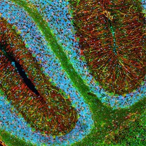
Only recognizes non-phosphorylated neurofilament H. By immunocytochemistry this antibody stains neuronal cell bodies, dendrites, and some thick axons in the central and peripheral nervous systems, but does not stain thin axons or other cells and tissues. Tissues and cultured cells can be fixed in a variety of paraformaldehyde- or formaldehye-containing fixatives, such as Bouin′s fixative. Antibody reactivity is poor in glutaraldehyde/paraformaldehyde-fixed samples. For staining formalin-fixed, paraffin sections it is recommended that de-paraffinized tissue be autoclaved in dH2O for 10 min or boiled in Tris-buffered saline, pH 9.0, in a microwave for 15 min. Trypsin pre-treatment abolishes antibody binding to the epitope. Post-fixation in cold methanol or methanol/hydrogen peroxide facilitates access of the antibody to the neurons in frozen sections or thick sections fixed in 4% paraformaldehyde and in cultured cells. By immunoblotting this antibody detects two bands, ~180 and ~200 kDa, which merge into a single NFH line on two-dimensional gels. Antibody should be titrated for optimal results in individual systems.
Trapp, B.D., et al. 1998. N. Engl. J. Med.338, 278.
King, C.E., et al. 1997. Neuroreport.8, 1663.
Campbell, M.J., et al. 1991. Brain Res.539, 133.
Campbell, M.J., et al. 1989. J. Comp. Neurol.282, 191.
Sternberger, L.A., et al. 1983. Proc. Natl. Acad. Sci. USA80, 6126.

Legal Information

CALBIOCHEM is a registered trademark of Merck KGaA, Darmstadt, Germany

Not finding the right product?  

Try our Product Selector Tool.

Regulatory Information

新产品

Certificates of Analysis (COA)

Search for Certificates of Analysis (COA) by entering the products Lot/Batch Number. Lot and Batch Numbers can be found on a product’s label following the words ‘Lot’ or ‘Batch’.

Already Own This Product?

Find documentation for the products that you have recently purchased in the Document Library.

Visit the Document Library

Danielle Beckman et al.
Proceedings of the National Academy of Sciences of the United States of America (2019-12-25)
As the average age of the population continues to rise, the number of individuals affected with age-related cognitive decline and Alzheimer's disease (AD) has increased and is projected to cost more than $290 billion in the United States in 2019.
Hana Yamate-Morgan et al.
Neuroscience, 406, 667-683 (2019-02-01)
Persistent demyelination has been implicated in axon damage and functional deficits underlying neurodegenerative diseases such as multiple sclerosis. The cuprizone diet model of demyelination allows for the investigation of mechanisms underlying timed and reproducible demyelination and remyelination. However, spontaneous oligodendrocyte
Joy Zhou et al.
eLife, 9 (2020-09-30)
Ramón y Cajal proclaimed the neuron doctrine based on circuit features he exemplified using cerebellar basket cell projections. Basket cells form dense inhibitory plexuses that wrap Purkinje cell somata and terminate as pinceaux at the initial segment of axons. Here
Milos D Ikonomovic et al.
Neuropharmacology, 145(Pt B), 160-176 (2018-06-23)
Traumatic brain injury (TBI) is a risk factor for development of chronic neurodegenerative disorders later in life. This review summarizes the current knowledge and concepts regarding the connection between long-term consequences of TBI and aging-associated neurodegenerative disorders including Alzheimer's disease
Lucia G Le Roux et al.
Cells, 9(5) (2020-05-28)
We illuminate a possible explanatory pathophysiologic mechanism for retinal cellular neuropathy by means of a novel diagnostic method using ophthalmoscopic imaging and a molecular imaging agent targeted to fast axonal transport. The retinal neuropathies are a group of diseases with

Our team of scientists has experience in all areas of research including Life Science, Material Science, Chemical Synthesis, Chromatography, Analytical and many others.

Contact Technical Service