Sign In to View Organizational & Contract Pricing.
Select a Size
About This Item
UNSPSC Code:
12352203
Clone:
SMI-311, monoclonal
Species reactivity:
-
Application:
—
Citations:
4
biological source
mouse
antibody form
ascites fluid
antibody product type
primary antibodies
clone
SMI-311, monoclonal
form
liquid
contains
≤0.1% sodium azide as preservative
species reactivity (predicted by homology)
mammals
manufacturer/tradename
Calbiochem®
storage condition
OK to freeze, avoid repeated freeze/thaw cycles
isotype
IgG1, IgGM
shipped in
wet ice
storage temp.
−20°C
target post-translational modification
unmodified
Quality Level
Gene Information
human ... NEFL(4747)
General description
Recognizes ~180-200 kDa pan-neuronal neurofilament marker in rat central nervous system cytoskeletal preparations.
This Anti-Pan-Neuronal Neurofilament Marker Mouse mAb (SMI-311) is validated for use in ELISA, Frozen Sections, WB, ICC, Paraffin Sections for the detection of Pan-Neuronal Neurofilament Marker.
Mouse monoclonal antibody supplied as undiluted ascites. Recognizes the ~180-200 kDa pan-neuronal neurofilament marker protein.
Immunogen
Rat
homogenized hypothalami extracted from Fischer 344 rat brain
Application

ELISA (1:1000)
Frozen Sections (1:1000, see comments)
Immunoblotting (1:1000)
Immunocytochemistry (1:1000, see comments)
Paraffin Sections (1:1000, heat pre-treatment required, see comments)
Physical form
Undiluted ascites.
Preparation Note
Following initial thaw, aliquot and freeze (-20°C).
Analysis Note
Positive Control
Rat brain or rat CNS cytoskeletal preparations
Rat brain or rat CNS cytoskeletal preparations
Other Notes
Agostino, A., et al. 2003. Hum. Mol. Genet.12, 399.
Tsunoda, I., et al. 2003. Am. J. Pathol.162, 1259.
Ulfig, N., et al. 1998. Cell Tissue Res.291, 433.
Tsunoda, I., et al. 2003. Am. J. Pathol.162, 1259.
Ulfig, N., et al. 1998. Cell Tissue Res.291, 433.
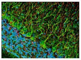
This antibody cocktail was selected to provide a specific marker for neurons in tissue sections and cultured cells. In contrast to individual anti-nonphosphoneurofilament antibodies that identify different subsets of neurons, this cocktail is a convenient marker for neurons in general and their differentiation from non-neuronal cells. Also useful as an early marker of neuronal migration and differentiation in human fetal development, yielding Golgi-like images without the disadvantages of the lack of selectivity and poor specificity of the Golgi technique. Can be used to trace the "inside-out gradient" of neuron production and differentiation in specifically delineating cell bodies and dendrites. Certain pathologic conditions, such as malnutrition, affect the SMI-311-visualized soma size and dendritic arborization. Tissues and cultured cells can be fixed in a variety of paraformaldehyde- or formaldehyde-containing fixatives, including Bouin′s fixative. This antibody does not react well with tissues or cells fixed in glutaraldehyde/paraformaldehyde. For staining paraffin sections it is recommended that de-paraffinized tissues be autoclaved in dH2O for 10 min to expose the epitope. Trypsin pre-treatment will abolish reactivity. Post-fixation in cold methanol or methanol/hydrogen peroxide facilitates access of the antibody to the neurons in frozen sections and thick tissues sections that have been fixed in 4% paraformaldehyde or cultured cells. For tissues that have not been paraffin-embedded and have been stored for long periods of time in formaldehyde fixatives, it is recommended that the tissues (up to 0.5 cm thick) be boiled in Tris-buffered saline, pH 9.0 for 15 min prior to sectioning. Antibody should be titrated for optimal results in individual systems.
Legal Information
CALBIOCHEM is a registered trademark of Merck KGaA, Darmstadt, Germany
Disclaimer
Toxicity: Standard Handling (A)
Not finding the right product?
Try our Product Selector Tool.
Storage Class
10 - Combustible liquids
wgk
WGK 1
Certificates of Analysis (COA)
Search for Certificates of Analysis (COA) by entering the products Lot/Batch Number. Lot and Batch Numbers can be found on a product’s label following the words ‘Lot’ or ‘Batch’.
Already Own This Product?
Find documentation for the products that you have recently purchased in the Document Library.
Timo Schomann et al.
Cell and tissue research, 381(1), 55-69 (2020-02-10)
Traumatic brain injury (TBI) is a devastating event for which current therapies are limited. Stem cell transplantation may lead to recovery of function via different mechanisms, such as cell replacement through differentiation, stimulation of angiogenesis and support to the microenvironment.
Yue Zhang et al.
The Journal of biological chemistry, 292(1), 100-111 (2016-11-30)
Astrocytes respond to CNS insults through reactive astrogliosis, but the underlying mechanisms are unclear. In this study, we show that inactivation of mechanistic target of rapamycin complex (mTORC1) signaling in postnatal neurons induces reactive astrogliosis in mice. Ablation of Raptor
Gabriela Rudd Garces et al.
Genes, 12(12) (2021-12-25)
We investigated a highly inbred family of British Shorthair cats in which two offspring were affected by deteriorating paraparesis due to complex skeletal malformations. Radiographs of both affected kittens revealed vertebral deformations with marked stenosis of the vertebral canal from
Hyun-Jeong Yang et al.
Nature communications, 7, 10884-10884 (2016-03-11)
While the formation of myelin by oligodendrocytes is critical for the function of the central nervous system, the molecular mechanism controlling oligodendrocyte differentiation remains largely unknown. Here we identify G protein-coupled receptor 37 (GPR37) as an inhibitor of late-stage oligodendrocyte
Our team of scientists has experience in all areas of research including Life Science, Material Science, Chemical Synthesis, Chromatography, Analytical and many others.
Contact Technical Service