Therapeutic Antibody Lead Discovery
Find the tools you need to select the most promising
lead candidates.
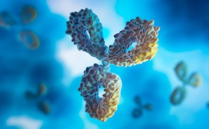
Therapeutic antibody lead discovery involves the selection of antibody candidates for targeted therapies. After a specific target has been identified, antibodies are produced using the target molecule as an antigen. Antibodies can be generated using methods including hybridoma technology, B cell cloning, and phage display. Antibodies are then purified and screened for their ability to bind antigen. Selected antibodies are further characterized to determine their specificity, affinity, stability, and pharmacokinetic properties. We provide numerous platforms and reagents to support the lead discovery process, from monoclonal antibody generation to isolation, selection, and lead optimization.
Lead Optimization for Monoclonal Antibodies
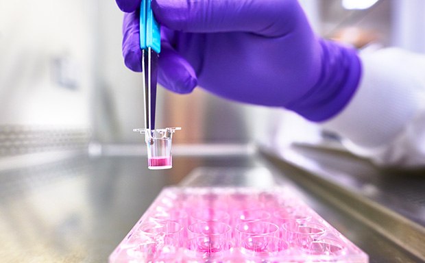
For optimized pharmacological drug efficacy, toxicity, and safety in monoclonal antibody drug development, we have an extensive portfolio of in vitro tools, cell models, and assays. Our innovative solutions include a broad range of cell lines and ready-to-assay cells for applications such as ADME (Absorption, Distribution, Metabolism, Excretion) assays and pharmacological studies. Our immuno-oncology panel cell lines provide an efficient and reliable model system for studying cancer immunotherapy. We also supply primary cells, PBMCs and NK cells, and patient-derived organoids for cellular models that more closely mimic the physiological environment and architecture of in vivo systems.
To support the development of safe and effective drug therapies, we have a range of cell health assays, including viability, apoptosis, and proliferation assays. Our live cell imaging reagents enable visualization and quantification of various cellular processes, providing insight into drug toxicity and efficacy.
Related Categories
ECACC repository cell lines, cancer cell lines, cellular models, and more.
Human primary cells, normal and disease state PBMCs, and immune cells.
Human iPSC-derived and patient-derived organoids for advanced cell culture.
Reagents, kits, and tools to measure general or specific indicators of cell status.
Fluorescent dyes, probes, biosensors, and labeling technologies for cell imaging.
Related Articles
- Cryopreserved PBMCs Retain Function and Phenotype
Frozen peripheral blood mononuclear cells offer many advantages over fresh PBMCs, enabling researchers to plan their experiments ahead of time with ready-to-use cells that can be stored in the freezer until needed.
- Gastrointestinal Organoid Biobank
Patient derived organoids (PDOs) can be used as in vitro 3D cell models, preserving original tissue physiology and molecular pathology for clinical relevance.
- Lung Cancer Organoid Biobank
Lung patient derived organoids (PDOs) have been shown to be a reliable model for lung cancer studies, and can also predict patient clinical responses to therapeutics.
- A Highly Sensitive IFN-γ ELISpot Assay to Quantify Cellular Immune Responses to Previous Viral Infection
This study presents an example of a typical ELISpot assay for quantifying the number of pre-existing, antigen-reactive T cells from peripheral blood mononuclear cell (PBMC) samples obtained from donors previously infected with cytomegalovirus (CMV).
- The ELISpot Assay Enables Functional Analysis of Cellular Immunology
Recent improvements to the design of multiwell microplates, including use of membranes with reduced background fluorescence, have bolstered the widespread application of ELISpot assays.
Explore our Products & Services
To continue reading please sign in or create an account.
Don't Have An Account?