Water for Nucleic Acid Gel Electrophoresis

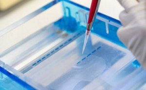
Loading of nucleic acid samples into an agarose gel for electrophoresis
What is Nucleic Acid Gel Electrophoresis?
Nucleic acid electrophoresis is the migration of DNA and RNA fragments under the influence of an electric field within a supporting matrix, called a gel. DNA and RNA fragments have a negative charge because of their phosphate backbone, and as such migrate toward the anode. The relative rate of migration of the fragments depends primarily on their net charge, size, and shape.
The gel, usually made of agarose or polyacrylamide, is immersed within an electrophoresis buffer that provides ions to carry a current and maintains the pH constant. The gel is porous and therefore acts as a sieve by retarding, or in some cases completely obstructing, the movement of large macromolecules while allowing smaller molecules to migrate freely.
The quality of the purified water used in the gels and TBE/TAE running buffers can affect the robustness and consistency of the band separation and signal intensity.
Types of gels used in nucleic acid electrophoresis
Agarose gels are commonly used for DNA and RNA electrophoresis. They have a large range of separation, but relatively low resolving power. By varying the concentration of agarose, fragments of DNA from about 50 to 50,000 base pairs (bp) can be separated using standard electrophoretic techniques.
Polyacrylamide gels have a rather small range of separation, but very high resolving power. In the case of DNA, polyacrylamide is used for separating fragments of less than about 500 bp. However, under appropriate conditions, fragments of DNA differing in length by a single base pair are easily resolved. These gels are also used extensively for separating and characterizing mixtures of proteins.
Staining nucleic acid gels
Once the gel has been run, the nucleic acids are stained and visualized, often with the fluorescent dye ethidium bromide (EtBr). EtBr intercalates with nucleic acid molecules and can be viewed under UV light. Fragment size determination is typically done by comparison to commercially available DNA ladders containing linear DNA fragments of known length.
Native vs. denatured gel electrophoresis
Native gels preserve the natural conformation of nucleic acids, separating them based on size, shape and charge. They are used when the native structure is important for analysis. Native gel electrophoresis is often used for studying nucleic acid-protein interactions, DNA or RNA folding structures, and for the purification of specific conformations of nucleic acids. It's also useful for separating supercoiled DNA from linear or open circular DNA.
In denatured gels, nucleic acids are linearized and denatured, allowing for separation purely based on size. This method is essential for applications requiring the analysis of precise nucleotide sequences or sizes. Denatured gel electrophoresis is crucial for sequencing, as it ensures that the DNA fragments are in a single-stranded form, which is necessary for precise size-based separation. It's also used in techniques like RNA analysis (including Northern blotting) where the RNA's secondary structure could affect the separation.
Southern and Northern Blotting
To identify the presence of a specific nucleotide sequence in a gel, Southern blotting (if DNA is being analyzed) or Northern blotting (RNA is being analyzed) is used. The DNA or RNA from the gel is transferred onto a nitrocellulose or nylon membrane. The membrane is then exposed to a hybridization probe—a single DNA fragment with a specific sequence whose presence in the target DNA (or RNA) is to be determined. The probe DNA is labeled so that it can be detected, usually by tagging with a fluorescent or chromogenic dye or sometimes incorporating radioactivity.
Impact of Water Quality on Nucleic Acid Electrophoresis
Nucleases (DNase, RNase)
DNA and RNA molecules analyzed by gel electrophoresis must stay intact. The presence of unwanted nucleases would degrade the nucleic acids in the sample, and the molecules of interest would not be detected. For the preparation of nuclease-free water, please refer to our technical article, Water for Nuclease-sensitive Molecular Biology Studies.
Ions
The ionic strength and pH of the buffers used to prepare the gel and the running buffers have to be constant. The presence of high levels of ions could alter the ionic strength of these solutions. High ionic strength can lead to excessive heat generation during electrophoresis, which can cause the gel to distort.1 Conversely, insufficient ionic strength might result in poor electrical conduction, leading to uneven migration of the nucleic acids. Fluctuations in pH can alter the charge on nucleic acid molecules, affecting their migration through the gel. A stable pH ensures consistent charge and migration patterns.
Metals
Certain metal ions, particularly divalent cations like Mg²⁺ and Ca²⁺, can facilitate the activity of nucleases that degrade nucleic acids. Trace metal contamination can thus result in the degradation of DNA or RNA during electrophoresis, appearing as smearing or degradation products on a gel.
Other metal ions can bind to the phosphate groups of nucleic acids, altering their overall charge and thus their mobility in the gel. For instance, heavy metals like lead or cadmium can bind to DNA and change its conformation, potentially leading to slower or irregular migration. In addition, some metals can catalyze undesirable chemical reactions in the gel. Iron, for example, can catalyze the formation of free radicals, which can break down both the gel matrix and the nucleic acids.2
Bacteria
Bacteria present in water could release degradation by-products such as the nucleases and ions previously mentioned and affect gel electrophoresis of nucleic acids.
Experiment to Assess the Impact of Water Quality on Nucleic Acid Electrophoresis
In an experiment, ribosomal RNA (rRNA) from E. coli was loaded onto an agarose gel. To assess the impact of water quality on the quality of electrophoresis results, the gels and buffers were prepared with either pure (Type 2) water or ultrapure (Type 1) water. Figure 1 shows that higher signal/band intensities were obtained in gels prepared and run with ultrapure water.

Figure 1.Agarose gel electrophoresis of rRNA. Gels and buffers were prepared using either (A) pure or (B) ultrapure water. The band intensities are higher with ultrapure water.
Ultrapure Water for Reliable and Consistent Nucleic Acid Electrophoresis
Ultrapure (Type 1) water purified with an ultrafiltration final filter is highly desirable for buffer and gel preparation in electrophoresis and related techniques to avoid contamination, improve sample separation, enhance signal intensity, and increase repeatability. Selecting the optimal buffer (e.g., TAE or TBE) and ensuring it is prepared with ultrapure water can help maintain consistent ionic strength and pH, reducing the risk of issues caused by ionic fluctuations. Using freshly purified ultrapure water with an ultrafilter at the point-of-use will ensure no bacteria or nucleases are present.
A range of water purification solutions adapted to the needs of scientists working with DNA and RNA separations is available.
Select and configure the optimal water purification system for your laboratory and its nucleic acid electrophoresis applications, or request support from a lab water solution expert.
Related Products
References
如要继续阅读,请登录或创建帐户。
暂无帐户?