In Vitro Differentiation of Human PBMC and CD34+ Derived Monocytes into Mature CD83+/CD14- Dendritic Cells
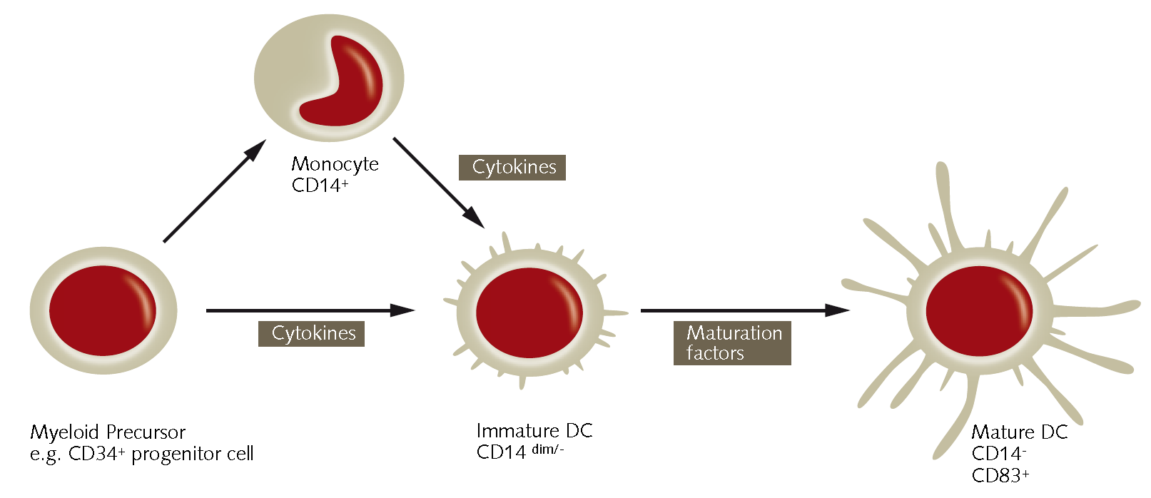
Dendritic Cells (DCs) are highly motile immune cells and are the most powerful antigen presenting cells (APCs) of the mammalian immune system.1 DC’s act as messengers between the innate and the adaptive immune systems by continuously sampling their environment for antigens by means of endocytosis.1 Being responsible for the induction T-cell dependent immunity and tolerance, they are especially abundant in epithelia (skin and intestinal tract) and other tissues that frquently encounter invading pathogens.2 Currently, there are two known major dendritic cell types in humans: 1) lymphoid dendritic cells derived from plasmacytoid cells in the blood and 2) myeloid dendritic cells derived from myeloid precursor cells such as peripheral blood monocytes (PBMCs) or CD34+ hematopoietic progenitor cells from the bone marrow. 1 DCs develop by means of a maturation process induced by exogenous or endogenous stimuli ( Fig.1). More recently, dendritic cells have been used for their potential development of cancer immunotherapies3 as well as the treatment of autoimmune diseases.4 Since DCs also play an essential role in the setting of HIV infection5 and the pathogenesis of several other viruses, they have significance as a therapeutical target.
PromoCell’s Dendritic Cell Generation Media (C-28050) allows for the efficient and reliable in vitro differentiation of human monocytes (hMo) into immature as well as fully mature CD83+/CD14- dendritic cells. The media can be used for the differentiation of cryopreserved purified monocytes into moDCs. Futhermore, the media may also be used with freshly isolated monocytes and MNCs when combined with the PromoCell Monocyte Attachment Medium (C-28051). The chemically defined and xeno-free PromoCell Dendritic Cell Generation Medium DXF (C-28052) is formualted exclusively with synthetic, recombinant or plant-sourced materials and is recommended for use with freshly isolated monocytes or mononuclear cells (MNCs).

Figure 1. Differentiation of human PBMC and CD34+ derived monocytes into mature CD83+/CD14-dendritic cells.
Dendritic Cell Differentiation Protocol
Protocol A: Generation of moDCs from freshly isolated cells using DC Generation Medium DXF
For Generation of moDCs from freshly isolated peripheral blood Monocytes or Mononuclear Cells, PromoCell recommends the use of the Dendritic Cell Generation Medium DXF (C-28052). Refer to protocol A for details.
Alternatively, the Dendritic Cell Generation Medium (C-28050) in combination with the Monocyte Attachment Medium (C-28051) may be used. See protocol B for detailed procedure.
- Let the cells attach (day 0). Plate freshly isolated cells in an appropriate amount of PromoCell DC Generation Medium DXF (C-28052) w/o cytokines. Mononuclear Cells at a density of 2-3 million/cm2 and purified monocytes at 0.5 million/cm2. Incubate for 1 hour at 5% CO2 and 37 °C in the incubator.
- Wash the adherent cell fraction (day 0). By vigorously swirling the tissue culture vessel, loosen non-adherent cells and aspirate them. Wash the adherent cells three times with warm PromoCell DC Generation Medium DXF (C-28052) w/o cytokines by swirling the vessel and aspirating the supernatant.
- Start differentiation into immature moDC (day 0). Add an appropriate amount of PromoCell DC Generation Medium DXF (C-28052) supplemented with 1x Component A of the Cytokine Pack moDC DXF (supplied at 100x) and incubate for 3 days at 37 °C and 5% CO2.
- Medium change (day 3). Perform a medium change on day 3: Aspirate the medium from the cells and collect it in a centrifugation tube. Immediately, pipet fresh PromoCell DC Generation Medium DXF (C-28052) supplemented with 1x Component A of the Cytokine Pack moDC DXF to the cells. Centrifuge the cells in the tube for 10 min at 180 x g. Discard the supernatant and carefully resuspend the cells in a small amount of fresh medium. Combine the resuspended cells in the tube with the cells in the fresh medium contained in the tissue culture vessel. Incubate the immature moDCs for another 3 days at 37 °C and 5% CO2.
- Complete moDC maturation process (day 6). To complete the moDC maturation process, supplement the whole volume with 1x of Component B of the Cytokine Pack moDC (supplied at 100x) on day 6. Do not change the medium. Incubate at 37 °C and 5% CO2 for an additional 24-48 hours.
- Harvest mature moDC (day 7/8). Dislodge loosely attached cells by pipetting up and down several times. Transfer the medium containing the cells in a 50 mL tube. Spin down harvested moDCs at 180 x g for 10 minutes and discard the supernatant.
- Perform your experiments. Resuspend and count the cells. The moDCs are now ready to be used in your experiments. Optionally, characterize their dendritic cell immuno-phenotype, e.g. by performing flow cytometry analysis for CD14, CD45 and CD83.
Protocol B: Generation of moDCs from freshly isolated cells using DC Generation Medium and Monocyte Attachment Medium
- Let the cells attach (day 0). Plate freshly isolated cells in an appropriate amount the PromoCell Monocyte Attachment Medium (C-28051). Plate mononuclear cells at a density of 2-3 million/cm2 and purified Monocytes at 0.5 million/cm2. Incubate for 1 hour at 5% CO2 and 37 °C in the incubator.
- Wash the adherent cell fraction (day 0). By vigorously swirling the tissue culture vessel, loosen non-adherent cells and aspirate them. Wash the adherent cells three times with warm Monocyte Attachment Medium (C-28051) by swirling the vessel and aspirating the supernatant.
- Start differentiation into immature moDC (day 0). Add an appropriate amount of PromoCell Dendritic Cell Generation Medium (C-28050) supplemented with 1x Component A of the Cytokine Pack moDC (supplied at 100x) and incubate for 3 days at 37 °C and 5% CO2.
- Medium Change (day 3). Perform a medium change on day 3: Aspirate the medium from the cells and collect it in a centrifugation tube. Immediately, pipet fresh PromoCell DC Generation Medium (C-28050) supplemented with 1x Component A of the Cytokine Pack moDC to the cells. Centrifuge the cells in the tube for 10 min at 180 x g. Discard the supernatant and carefully resuspend the cells in a small amount of fresh medium. Combine the resuspended cells in the tube with the cells in the fresh medium contained in the tissue culture vessel. Incubate the immature moDCs for another 3 days at 37 °C and 5% CO2.
- Complete moDC maturation process (day 6). To complete the moDC maturation process, supplement the whole volume with 1x of Component B of the Cytokine Pack moDC (supplied at 100x) on day 6. Do not change the medium. Incubate at 37 °C and 5% CO2 for an additional 24-48 hours.
- Harvest mature moDC (day 7/8). Dislodge loosely attached cells by pipetting up and down several times. Transfer the medium containing the cells in a 50 mL tube. Spin down harvested moDCs at 180 x g for 10 minutes and discard the supernatant.
- Perform your experiments. Resuspend and count the cells. The moDCs are now ready to be used in your experiments. Optionally, characterize their dendritic cell immuno-phenotype, e.g. by performing flow cytometry analysis for CD14, CD45 and CD83.
Protocol C: Generation of moDCs from Cryopreserved Cells
For Generation of moDCs from cryopreserved peripheral blood Monocytes, PromoCell recommends the use of the Dendritic Cell Generation Medium (C-28050).
- Plate the cells (day 0). Thaw cryopreserved monocytes in a waterbath according to the instruction manual delivered with the cells. After thawing, immediately plate them at 0.5 million/cm2 in an appropriate amount of PromoCell Dendritic Cell Generation Medium (C-28050) supplemented with 1x Component A of the Cytokine Pack moDC (supplied at 100x). Use at least 9 mL medium per vial of cryopreserved cells. Immediately place them in an incubator for 1 day at 37 °C and 5% CO2.
- Medium change (day 1). Aspirate the medium from the cells and collect it in a centrifugation tube. Immediately pipet fresh PromoCell Dendritic Cell Generation Medium (C-28050) supplemented with 1x Component A of the Cytokine Pack moDC to the cells. Centrifuge the cells in the tube for 10 min at 180 x g. Discard the supernatant and carefully resuspend the cells in a small amount of fresh medium. Combine the resuspended cells with the cells in the fresh medium contained in the tissue culture vessel. Incubate for 3 more days.
- Medium change (day 4). Perform a medium change as described above. Incubate for a further 2 days at 37 °C and 5% CO2.
- Complete moDC maturation process (day 6). To complete the moDC maturation process, supplement the whole volume with 1x of Component B of the Cytokine Pack moDC (supplied at 100x) on day 6. Do not change the medium. Incubate at 37 °C and 5% CO2 for an additional 24 to 48 hours.
- Harvest mature moDC (day 7/8). Dislodge loosely attached cells by pipetting up and down several times. Transfer the medium containing the cells to a 50 mL tube. Spin down harvested moDCs at 180 x g for 10 minutes and discard the supernatant.
- Perform your experiments. Resuspend and count the cells. The moDC are now ready to be used in your experiments. Optionally, characterize their dendritic cell immuno-phenotype, e.g. by performing flow cytometry analysis for CD14, CD45 and CD83.
Results
A B


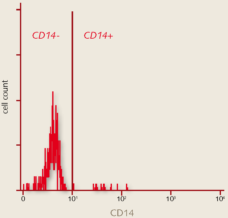
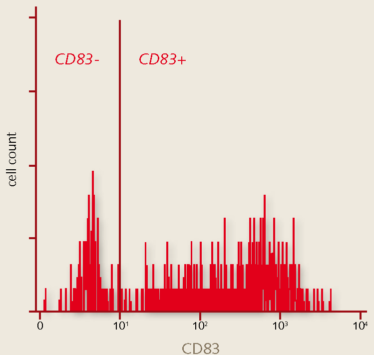
Figure 2. FACS analysis of mature monocyte derived human dendritic cells. Mature dendritic cell markers include high expression of CD83 and low expression of CD14. A. Flow-cytometry analysis of day 8 mature moDCs generated in the PromoCell DC Generation Medium are negative for the immature marker CD14 and positive for the DC maturation marker CD83.

Figure 3. Morphology of immature and mature monocyte derived human dendritic cells. A) Day 6 immature monocyte-derived dendritic cells (moDCs). Note the large cytoplasmic, veil-like processes seen in adherent (center) as well as loosely attached (upper left) cells. B) Day 8 mature monocyte-derived Dendritic Cell (moDC). Note the multiple dendrite-like structures arising from the surface of this non-adherent cell.
Materials
References
To continue reading please sign in or create an account.
Don't Have An Account?