Visikol® HISTO™ Tissue Clearing Reagents
One of the most impactful trends to occur recently in histopathology is the popularization of new tissue clearing methods in microscopy and high-content applications. These techniques render tissues transparent by harmonizing the mismatched refractive indices of cellular components, enabling true 3D microscopic analysis of intact biological structures, rather than prevailing methods that required the laborious preparation and analysis of individual image slices. The power of tissue clearing has already been harnessed by leading researchers to address key questions at the frontiers of neuroscience, cancer biology, and developmental biology. With the multitude of tissue clearing methods available today, it is important to understand how aqueous and solvent-based approaches impact the output of the data, the assay workflow and the tissue specimen itself. We are partnering with Visikol® to offer the HISTO™ and HISTO-M™ reagents, which can be used to easily and reversibly clear tissue or 3D cell cultures while retaining sample integrity for downstream analysis.
Visikol® tissue clearing method features and benefits include:
- Fast, non-toxic workflow with no special equipment or acrylamide embedding
- Effectively perform immunostaining or fluorescent protein analysis on cleared samples
- Retain morphology and lipid content information in your samples
- A reversible clearing procedure to conduct additional downstream analysis
- Access to proven protocols for a variety of tissue and cell imaging applications

Figure 1.Visikol® Histo™ reagent tissue clearing

Figure 2. A, B.Precision cut lung slice before (left) and after (right) subjected to tissue clearing by Visikol method HepG2 spheroids stained with Calcein AM, DAPI, and PI.
Visikol® HISTO™ 3D tissue clearing
Traditional tissue clearing methods are often limited by the non-reversible nature of the clearing reagent and the overall disruption in the morphology of the cells and tissue themselves. Tissue clearing methods for 3D cell culture and tissue samples are increasingly being used by researchers to improve models in use for both fundamental biology and drug discovery research. Visikol® HISTO™ 3D tissue clearing technology opens the possibility for cell culture and tissue models that deliver qualitative and quantitative data. With a fully reversible tissue clearing protocol, researchers can generate thorough data sets of the highest quality while still maintaining the integrity of their experimental samples.

Reversible nature of HISTO™ tissue clearing method
Left panel: rat brain after clearing
Right panel: tissue opacity restored after reversal.


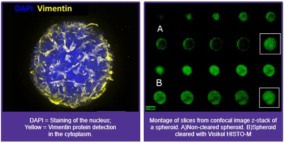
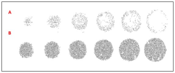
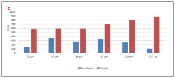
Nuclei stained with SYTOX green; A) Non-cleared spheroid; B) Cleared spheroid; Z-projections are outlined in white. Z-projections mask difficulty of imaging non-cleared microtissues since Z-projections hide the eclipsing that occurs in imaging. Cleared microtissues show no eclipsing while interior cells are visible. Outlines of cells detected by CellProfiler in InSphero NCI-H2170 lung cancer microtissues treated with 100 nM cisplatin. 20 µm optical-sections (20-120 um) shown left to right for A) Non-cleared microtissues and B) Microtissues cleared with Visikol® HISTO-M™; C) Cell counts for each Z-plane in cleared and non-cleared microtissues from A) and B). Far more cells can be detected at each Z-plane in cleared microtissues.

Visikol® HISTO™ for drug discovery
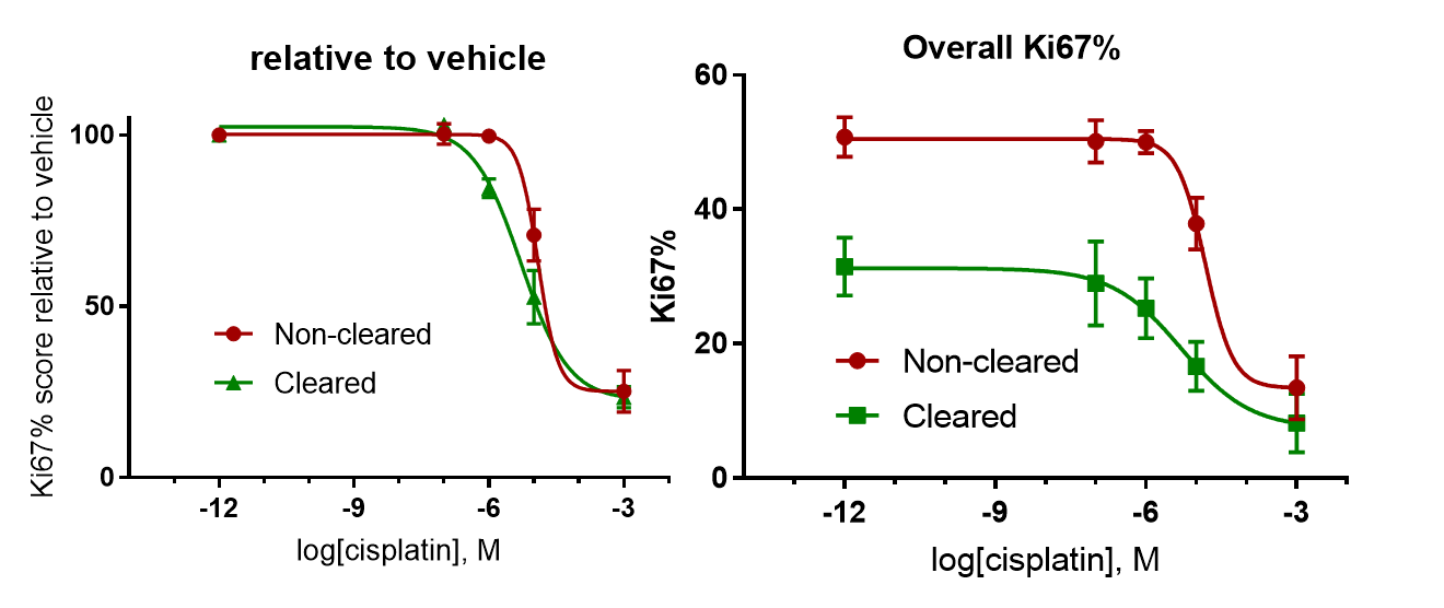
Antiproliferation dose response curves for cisplatin-treated NCI-H2170 microtissues A) relative to vehicle control cell proliferation score; B) showing absolute Ki67% proliferation score.
Materials
To continue reading please sign in or create an account.
Don't Have An Account?