Immunoprecipitation of FLAG Fusion Proteins Using Monoclonal Antibody Affinity Gels
- What is the FLAG peptide tag, and how is it used to isolate and purify proteins?
- Immunoprecipitation Technical Overview
- Protocol for Immunoprecipitation (IP) of FLAG Fusion Proteins Using M2 Affinity Gels
- Purification of Small Amounts of FLAG Fusion Proteins
Immunoprecipitation (IP) can be used for efficient, high-yield isolation and purification of proteins fused to the FLAG® peptide tag. IP is performed with the ANTI-FLAG® M2 affinity gel, which is a highly specific monoclonal antibody covalently bound to agarose resin. Affinity resin permits efficient binding of FLAG®-tagged proteins without the need for preliminary steps and calibrations. The immunoprecipitated FLAG®-tagged fusion proteins can be efficiently eluted from the resin with acidic conditions or by competition with FLAG® peptide. The immunoprecipitated proteins can then be analyzed for their size, post-translational modifications, and gel interactions during electrophoresis, as well as by activity assays .
What is the FLAG peptide tag, and how is it used to isolate and purify proteins?
FLAG® peptides are marker peptides used in protein detection and purification. The FLAG® tag peptide is composed of eight amino acids (DYKDDDDK) that maximize immunogenicity, so that high-affinity monoclonal antibodies raised against the FLAG® sequence can be used to detect and isolate proteins containing the FLAG® peptide. Using recombinant technique, FLAG®marker peptides can be added at the protein N-terminus, the C-terminus, or the N-terminus preceded by a methionine residue (Met-FLAG®), and in internal positions
Immunoprecipitation Technical Overview
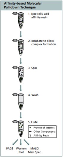
Immunoprecipitation consists of the following steps and intermediates: cell lysis, binding of specific antigen to an antibody, antibody-antigen complex, precipitation, precipitant wash, and antigen dissociation from the immune complex.

Protocol for Immunoprecipitation (IP) of FLAG Fusion Proteins Using M2 Affinity Gels Which Contain M2 Monoclonal Antibody Bound Covalently to Crosslinked 4% Agarose Beads
Use EZ view red anti-FLAG M2 affinity gel F2426, or Anti-FLAG M2 affinity gel A2220.
Purification of Small Amounts of FLAG Fusion Proteins
Note: Two control reactions are recommended for the immunoprecipitation procedure
- Positive control (FLAG-BAP fusion protein, like the Amino-terminal P7582, Carboxy-terminal P7457 or Met-FLAG-BAP fusion protein P5975)
- Negative control (reagent blank with NO protein)
a) Carefully mix the affinity gel beads until completely suspended. Immediately aliquot 40 µL of the 50% slurry (20 µL of packed gel volume) into a 1.5 mL microcentrifuge tube using a wide orifice pipette tip, or cut 1 mm off the end of a regular pipette tip.
b) Wash/equilibrate beads: Add 500 µL of TBS (50 mM Tris HCL, 150 mM NaCl, pH 7.4), vortex and centrifuge for 30 – 60 seconds at 5000-8,200 x g. Remove supernatant, being careful not to disturb the beads.
c) Wash again as above, and set the tube on ice until sample is ready to add. Note: When multiple immunoprecipitation samples are processed simultaneously, the combined amount of resin needed can be washed together. Each wash should be performed with TBS at a volume of at least 20 times the total packed gel volume. Once washed, the resin can be aliquoted for the desired number of samples.
d) Sample preparation:
- Clarify the cell lysate by centrifugation (14,000 x g for 10–15 min at 2–4°C)
- Calculate the amount of lysate needed. Volume will depend on the expression level of the FLAG fusion protein in the transfected cells.
e) Bind: Add 200–1000 µL of cell lysate (if necessary, bring final volume to 1 mL by adding lysis buffer [50 mM Tris HCl, pH 7.4, 150 mM NaCl, 1 mM EDTA, 1% Triton X-100]). Note: volume of lysate will be dependent on the expression level of FLAG fusion protein in the transfected cells. For positive control, add 1 mL of TBS and 4 µL of 50 ng/µL of FLAG-BAP fusion protein (about 200 ng). For negative control add 1 mL of lysis buffer only. Incubate all samples and controls with shaking using a roller shaker or end-over-end shaker for about 1–2 hours at 2–8°C (binding step can be extended overnight to ensure maximum binding).
f) Centrifuge the tube for 30 sec–1 min at 5000- 8,200 x g. and aspirate the supernatant (flow-through). Flowthrough may be retained if desired.
g) Wash the beads: Add 500 µL of TBS, vortex and centrifuge for 30 sec–1 min at 5000- 8,200 x g. Remove supernatant
h) Repeat wash two more times.
i) Elution of the sample: Three elution methods can be performed, and the choice will depend on characteristics of the tagged protein, as well as the downstream application.
- Elution of the FLAG fusion protein may be achieved under native conditions by competition using 3X FLAG peptide (F4799). This elution is the most efficient method.
- Elution may be performed under acidic conditions using 0.1 M glycine HCl, pH 3.5. This elution method is fast and efficient, but requires immediate neutralization of the sample.
- Elution may be achieved by electrophoresis using SDS-PAGE sample buffer. If this method is employed, the beads cannot be used again since SDS will denature the M2 antibody.
Elution with 3X FLAG Peptide (F4799)
i. Preparation of the 3X FLAG peptide:
- Dissolve the 3X FLAG peptide in 0.5 M Tris HCL, pH 7.5 1 M NaCl at final concentration of 25 µg/µL
- Dilute the stock solution 5-fold with diH20 to a final concentration of 5 µg/µL
- Prepare the Elution working solution at 150 ng/µL, by adding 3 µL of the 5 µg/µL stock to 100 µL of TBS.
ii. Add 100 μL of 3X FLAG elution working solution to each sample and incubate for 30 min at 2–8°C with gentle shaking.
iii. Centrifuge for 30 sec–1 min at 5000-8,200 x g. Transfer the supernatant to a fresh tube. Be careful not to transfer any resin.
1. Elution under acidic conditions (0.1 M glycine HCl pH -3.5) Procedure performed at room temperature.
i. Add 100 µL of 0.1 M glycine HCl, pH 3.5, to each sample
ii. Incubate for 5–10 min with gentle shaking
iii. Centrifuge the resin for 30 sec–1 min at 5000-8200 x g
iv. Neutralize the sample by transfer of the supernatant to fresh tubes containing 10 µL of 0.5 M Tris HCl, pH 7.4, with 1.5 M NaCl
v. Use immediately or store at -20°C for long term.
2. Elution with SDS-PAGE sample buffer (62.5mM Tris HCl, pH 6.8 with 2% SDS, 10% (v/v) glycerol and 0.002% bromophenol blue. Procedure performed at room temperature.
Note: In order to minimize the denaturation of antibody, no reducing agent should be included (DTT or 2-mercaptoethanol). Addition of reducing agents will dissociate the heavy and light chains of the immobilized M2 antibody (25–50kDa bands)
i. Add 20 µL of 2X sample buffer to each sample
ii. Boil the samples for 3 min
iii. Centrifuge for 30 sec – 1 min at 5000-8200 x g
iv. Transfer supernatant to fresh tube. Samples are ready for loading on SDS-PAGE.

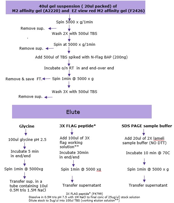
Figure 1.Summary of the options used for Immunoprecipitation in batch mode for low abundance proteins
Materials
To continue reading please sign in or create an account.
Don't Have An Account?